. 2025 Jun 24;13(7):1466.
doi: 10.3390/microorganisms13071466.

Germinated Spores of the Probiotic Bacterium Bacillus coagulans JBI-YZ6.3 Support Dynamic Changes in Intestinal Epithelial Communication and Resilience to Mechanical Wounding

Affiliations

Germinated Spores of the Probiotic Bacterium Bacillus coagulans JBI-YZ6.3 Support Dynamic Changes in Intestinal Epithelial Communication and Resilience to Mechanical Wounding

Sage V McGarry et al. Microorganisms. .

Abstract

The spore-forming probiotic Bacillus coagulans JBI-YZ6.3 interacts with the gut epithelium via its secreted metabolites as well as its cell walls, engaging pattern-recognition receptors on the epithelium. We evaluated its effects on human T84 gut epithelial cells using in vitro co-cultures, comparing metabolically active germinated spores to the isolated metabolite fraction and cell wall fraction under unstressed versus inflamed conditions. Germinated spores affected epithelial communication via chemokines interleukin-8, interferon gamma-induced protein-10, and macrophage inflammatory protein-1 alpha and beta after 2 and 24 h of co-culture. Non-linear dose responses confirmed that bacterial density affected the epigenetic state of the epithelial cells. In contrast, the cell wall fraction increased cytokine and chemokine levels under both normal and inflamed conditions, demonstrating that the intact bacterium had anti-inflammatory properties, regulating pro-inflammatory signals from its cell walls. During recovery from mechanical wounding, germinated spores accelerated healing, both in the absence and presence of LPS-induced inflammation; both the metabolite and cell wall fractions contributed to this effect. The release of zonulin, a regulator of tight junction integrity, was reduced by germinated spores after 2 h. These findings suggest that B. coagulans JBI-YZ6.3 modulates epithelial chemokine signaling, supports barrier integrity, and enhances epithelial resilience, highlighting its potential as an efficacious multi-faceted probiotic for gut health.

Keywords: IL-6; IL-8; IP-10; MIP-1α; MIP-1β; anti-inflammatory; chemotactic recruitment; wound healing; zonulin.


Conflict of interest statement

The authors declare no conflicts of interest. All authors are affiliated with NIS Labs. Jeneil Biotech Inc. sponsored the research for this work to be performed at NIS Labs. The sponsor had no role in the design of the study; in the collection, analyses, or interpretation of data; in the writing of the manuscript; or in the decision to publish the results.

Figures

Figure 1
Figure 1
Diagram illustrating the gut epithelial cell layer, separating the gut lumen from the underlying tissues. The epithelial cells play pivotal roles in communication between the gut microbiome and the host.
Figure 2
Figure 2
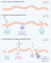
Gut epithelial structure and function. Zonulin plays an active regulating role in tight junctions. (A) Static epithelial barrier. (B) Healthy epithelial barrier, where dynamic changes to tight junctions allow paracellular elimination of invading bacteria, immune cell scavenging of luminal antigens, and paracellular transport of nutrients. (C) Pathologically disrupted barrier where nutrients are leaking, and opportunistic bacteria penetrate across the barrier. Allergens (environmental and food-related) can cross the barrier. Both bacterial and allergen antigens can elicit inappropriate immune responses by presentation to dendritic cells out of context.
Figure 3
Figure 3
Relative metabolic activity of the germinated spores of B. coagulans JBI-YZ6.3 in the absence versus presence of penicillin, streptomycin, and fetal calf serum (FCS), mimicking the cell culture conditions used for the experimental testing in co-cultures of B. coagulans with T84 human gut epithelial cells. At 24 h, the metabolic activity of B. coagulans treated with antibiotics was statistically significantly lower than the cultures without antibiotics (p < 0.05). For the 1:40 and 1:160 dilutions, the metabolic activity of cultures in the presence of fetal calf serum was significantly higher than that of cultures treated with penicillin and streptomycin in the absence of fetal calf serum (p < 0.01).
Figure 4
Figure 4
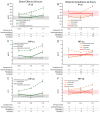
Changes to IL-6 and IL-8 levels in T84 cell cultures after 2 h of exposure to germinated spores, metabolite fraction, and cell wall fraction. The direct effects under normal culture conditions are shown in panels (A,C). The effects in the context of LPS-induced inflamed conditions are shown in panels (B,D). The data are shown as the averages ± standard deviation of duplicate data points, compared to control cultures as hexaplicate data points. The averages of untreated control cultures are shown as gray lines, and the standard deviation as a gray shaded area. For IL-6, the lowest dose of the germinated spores had only one data point (not duplicate), and the statistical significance was not calculable (NC); the remaining 2 h data for IL-6 were below levels of significance and also not calculable (NC). The averages of LPS-treated control cultures are shown as red lines, and the standard deviation as a red shaded area. Where the standard deviation is “0”, no shaded area is visible. Levels of statistical significance are shown when comparing the test products to controls, indicated with (*) for p < 0.1 and * for p < 0.05.
Figure 5
Figure 5
Changes to chemokine levels in T84 cell cultures after 2 h of exposure to germinated spores, metabolite fraction, and cell wall fraction. The direct effects under normal culture conditions are shown in panels (A,C,E). The effects in the context of LPS-induced inflamed conditions are shown in panels (B,D,F). The data are shown as the averages ± standard deviation of duplicate data points, compared to control cultures as hexaplicate data points. The averages of untreated control cultures are shown as gray lines, and the standard deviation as a gray shaded area. For the lowest dose of the germinated spores, only a single data point was available (not duplicate), and the levels of significance were not calculable (NC). Levels of statistical significance are shown when comparing the test products to controls, indicated with (*) for p < 0.1, * for p < 0.05, and ** for p < 0.01.
Figure 6
Figure 6
Changes to IL-6 and IL-8 levels in T84 cell cultures after 24 h of exposure to germinated spores, metabolite fraction, and cell wall fraction. The direct effects under normal culture conditions are shown in panels (A,C). The effects in the context of LPS-induced inflamed conditions are shown in panels (B,D). The data are shown as the averages ± standard deviation of duplicate data points, compared to control cultures as hexaplicate data points. The averages of untreated control cultures are shown as gray lines, and the standard deviation as a gray shaded area. The averages of LPS-treated control cultures are shown as red lines, and the standard deviation as a red shaded area. Levels of statistical significance are shown when comparing the test products to controls, indicated with (*) for p < 0.1, * for p < 0.05, and ** for p < 0.01.
Figure 7
Figure 7
Changes to chemokine levels in T84 cell cultures after 24 h of exposure to germinated spores, metabolite fraction, and cell wall fraction. The direct effects under normal culture conditions are shown in panels (A,C,E). The effects in the context of LPS-induced inflamed conditions are shown in panels (B,D,F). The data are shown as the averages ± standard deviation of duplicate data points, compared to control cultures as hexaplicate data points. The averages of untreated control cultures are shown as gray lines, and the standard deviation as a gray shaded area. The averages of LPS-treated control cultures are shown as red lines, and the standard deviation as red shaded areas. Levels of statistical significance are shown when comparing the test products to controls, indicated with (*) for p < 0.1, * for p < 0.05, and ** for p < 0.01.
Figure 8
Figure 8
Wound healing after a mechanical scratch under normal (A) and LPS-induced inflamed (B) culture conditions. Results are shown as averages ± standard deviations for triplicate data points for each dose of germinated spores, metabolite fraction, cell wall fraction, and for each negative and positive control. The average of untreated control cultures is shown as a gray line, and the standard deviation as a gray shaded area. The average of LPS-treated control cultures is shown as a red line, and the standard deviation as a red shaded area. Levels of statistical significance are shown when comparing the test products to controls, indicated with (*) for p < 0.1 and * for p < 0.05.
Figure 9
Figure 9
Zonulin release after (A) 2 h and (B) 24 h of exposure to the probiotic test products. Results are shown as averages ± standard deviations for triplicate data points for germinated spores, the metabolite fraction, and the cell wall fraction. The average of untreated control cultures is shown as a gray line, and the standard deviation as a gray shaded area. Levels of statistical significance are shown when comparing the test products to the untreated controls, indicated with (*) for p < 0.1, * for p < 0.05, and ** for p < 0.01.
Figure 10
Figure 10
Schematic diagram illustrating the possibility of quorum sensing in relation to the probiotic bacterial density in the co-cultures of T84 gut epithelial cells with B. coagulans. At lower bacterial densities, planktonic forms of B. coagulans may have produced autoinducers capable of triggering altered gene expression in the T84 cells. In contrast, at the highest dose, B. coagulans may have formed biofilms on the apical side of the T84 cells, resulting in a different epigenetic state and a different profile of secreted metabolites affecting the T84 cells.

Similar articles

References

    1. Hrncir T. Gut Microbiota Dysbiosis: Triggers, Consequences, Diagnostic and Therapeutic Options. Microorganisms. 2022;7:578. doi: 10.3390/microorganisms10030578. - DOI - PMC - PubMed
    1. Wardman J.F., Bains R.K., Rahfeld P., Withers S.G. Carbohydrate-active enzymes (CAZymes) in the gut microbiome. Nat. Rev. Microbiol. 2022;20:542–556. doi: 10.1038/s41579-022-00712-1. - DOI - PubMed
    1. Oliphant K., Allen-Vercoe E. Macronutrient metabolism by the human gut microbiome: Major fermentation by-products and their impact on host health. Microbiome. 2019;7:91. doi: 10.1186/s40168-019-0704-8. - DOI - PMC - PubMed
    1. Fan P., Li L., Rezaei A., Eslamfam S., Che D., Ma X. Metabolites of Dietary Protein and Peptides by Intestinal Microbes and their Impacts on Gut. Curr. Protein Pept. Sci. 2015;16:646–654. doi: 10.2174/1389203716666150630133657. - DOI - PubMed
    1. McDermott A.J., Huffnagle G.B. The microbiome and regulation of mucosal immunity. Immunology. 2014;142:24–31. doi: 10.1111/imm.12231. - DOI - PMC - PubMed

Grants and funding

LinkOut - more resources