. 2025 Jun 24;18(7):950.
doi: 10.3390/ph18070950.

Resveratrol Alleviates Inflammatory Response Through P2X7/NLRP3 Signaling Pathway: In Silico and In Vitro Evidence from Activated Microglia

Affiliations

Resveratrol Alleviates Inflammatory Response Through P2X7/NLRP3 Signaling Pathway: In Silico and In Vitro Evidence from Activated Microglia

Bianca Fagan Bissacotti et al. Pharmaceuticals (Basel). .

Abstract

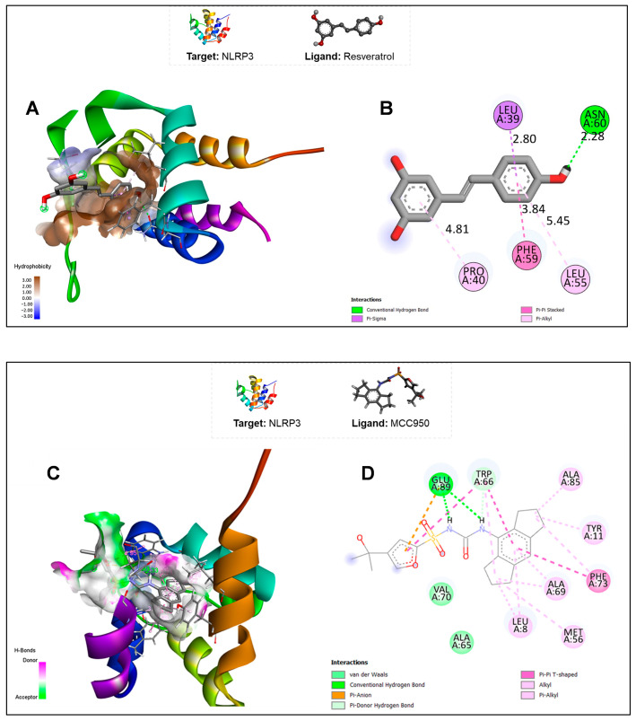
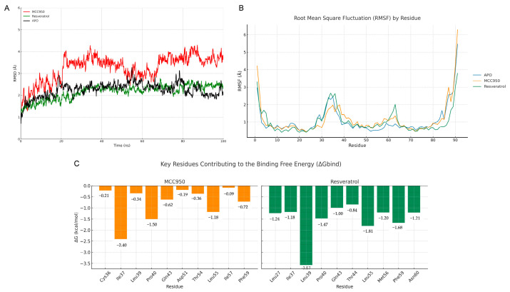
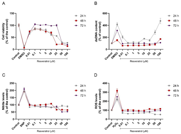
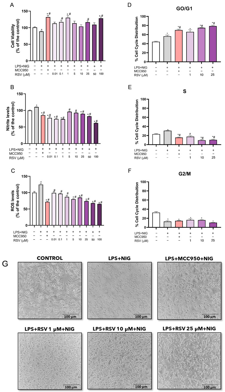
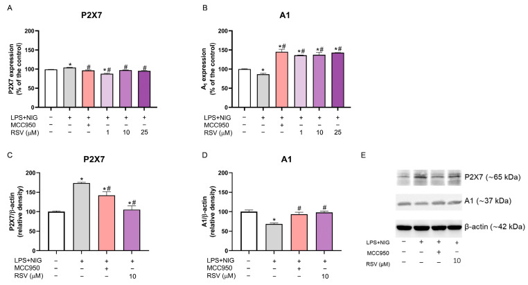
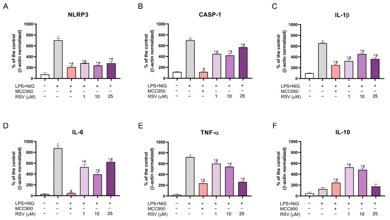
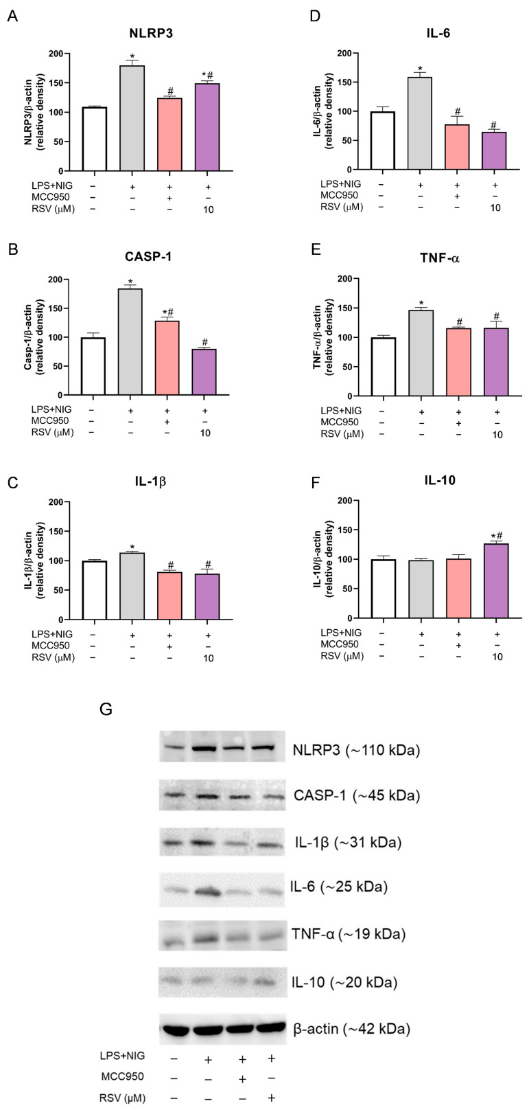
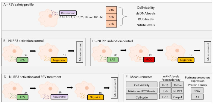
Background/Objectives: Chronic inflammation and inappropriate NLRP3 inflammasome regulation are related to many brain diseases. Purinergic mediators may play an important role in inflammation regulation and could be targeted for effective therapies for these illnesses. We evaluated resveratrol's anti-neuroinflammatory potential in BV-2 microglia cells using an innovative in vitro method of NLRP3 inflammasome activation, correlating with the P2X7 purinergic receptor. Methods: In silico analyses were used to estimate resveratrol's interaction with NLRP3, and its cytotoxicity was measured for 24, 48, and 72 h. Moreover, microglia were exposed to lipopolysaccharide and nigericin to activate the NLRP3 inflammasome and treated with resveratrol between these inflammatory agents. Results: It was found that resveratrol has binding compatible with modulating NLRP3. Specifically, 0.1-25 µM of resveratrol presented a favorable safety profile in BV-2 cells. Microglia exposed to the inflammatory agents had increased levels of oxidative species, the P2X7 receptor, and pro-inflammatory cytokines. However, resveratrol decreased the NLRP3, caspase-1, IL-1β, IL-6, and TNF-α mRNA levels and protein density; on the other hand, IL-10 was increased, acting as a protector, preventing exacerbated inflammation. Under resveratrol exposure, P2X7 was negatively expressed, regulating inflammation to establish homeostasis and microglial proliferation. Additionally, resveratrol activates the A1 adenosine receptor, possibly correlated with neuroprotective effects. Conclusions: We confirmed the anti-neuroinflammatory action of resveratrol via the P2X7 receptor and NLRP3's combined modulation, regulating the cell cycle and reducing pro-inflammatory and oxidant agents. Considering this pathway, resveratrol could be a candidate for further investigations as a potential treatment against neuroinflammatory diseases.

Keywords: BV-2 cells; brain inflammation; immunomodulation; polyphenol; purinergic receptors.


Conflict of interest statement

The authors declare no conflicts of interest.

Figures

Figure 1
Figure 1
(A) Interactions between the PYD domain of the NLRP3 protein and RSV, highlighting its hydrophobicity; (B) 2D map of interactions between the PYD domain of the NLRP3 protein and RSV, highlighting the amino acid residues that participate in the bonds and the types of bonds; (C) interactions between the PYD domain of the NLRP3 protein and MCC950, highlighting the regions of hydrogen bond formation; (D) 2D map of interactions between the PYD domain of the NLRP3 protein and MCC950, highlighting the amino acids that participate in the bonds and the types of bonds.
Figure 2
Figure 2
(A) Root mean square deviation (RMSD) of the simulations for the APO, MCC950, and RSV systems; (B) root mean square fluctuation (RMSF) per residue for the APO, MCC950, and RSV systems; (C) per-residue binding free energy contributions of MCC950 and RSV to the PYD domain (PDB ID: 2NAQ).
Figure 3
Figure 3
Concentration–response curve of RSV in the microglial cells (BV-2 cell line)—in vitro safety profile. (A) Cell viability for 24, 48, and 72 h exposure to RSV; (B) dsDNA free content for 24, 48, and 72 h exposure to RSV; (C) nitrite levels for 24, 48, and 72 h exposure to RSV; (D) ROS levels for 24, 48, and 72 h exposure to RSV. SNP—sodium nitroprusside. The experiments were performed in triplicate. The statistical analysis is demonstrated in Supplementary Material S1 (with the same results in a column graph) and was conducted by one-way ANOVA followed by the Tukey post hoc test. Data are expressed as mean values ± SEM.
Figure 4
Figure 4
Effects of RSV on cell viability, oxidative profile, and cell cycle of BV-2 cells subjected to the NLRP3 activation protocol. (A) Cell viability; (B) nitrite levels; (C) ROS levels; (DF) phases of cell cycle; (G) illustrative image of BV-2 cells and their respective treatments. Scale bar: 100 μm. The experiments were performed in triplicate. The statistical analysis was conducted using one-way ANOVA followed by the Tukey post hoc test. p < 0.05 was considered significant. Data are expressed as mean values ± SEM. * means comparison to the control group; # means comparisons to the activation group (LPS + NIG).
Figure 5
Figure 5
Effects of RSV on the purinergic receptor in BV-2 cells subjected to the NLRP3 activation protocol. (A) P2X7 expression by flow cytometer analysis; (B) A1 expression by flow cytometer analysis; (C) relative protein density of P2X7 receptor by Western blot analysis; (D) relative protein density of A1 receptor by Western blot analysis; (E) Western blot analysis showing the respective protein bands. The experiments were performed in triplicate. The statistical analysis was conducted using one-way ANOVA followed by the Tukey post hoc test. p < 0.05 was considered significant. Data are expressed as mean values ± SEM. * means comparison to the control group; # means comparisons to the activation group (LPS + NIG).
Figure 6
Figure 6
RSV modulation in the inflammatory mRNA levels in BV-2 microglial cells under exposure to NLRP3 inflammasome activation conditions. (A) NLRP3 mRNA levels; (B) caspase-1 mRNA levels; (C) IL-1β mRNA levels; (D) IL-6 mRNA levels; (E) TNF-α mRNA levels; (F) IL-10 mRNA levels. The experiments were performed in triplicate. The statistical analysis was conducted using one-way ANOVA followed by the Tukey post hoc test. p < 0.05 was considered significant. Data are expressed as mean values ± SEM. * means comparison to the control group; # means comparisons to the activation group (LPS + NIG).
Figure 7
Figure 7
Western blot analysis shows RSV modulation in the relative protein density of inflammatory markers. (A) Relative protein density of NLRP3; (B) relative protein density of caspase-1; (C) relative protein density of IL-1β; (D) relative protein density of IL-6; (E) relative protein density of TNF-α; (F) relative protein density of IL-10; (G) Western blot analysis showing the respective protein bands. The experiments were performed in triplicate. The statistical analysis was conducted using one-way ANOVA followed by the Tukey post hoc test. p < 0.05 was considered significant. Data are expressed as mean values ± SEM. * means comparison to the control group; # means comparisons to the activation group (LPS + NIG).
Figure 8
Figure 8
An illustrated scheme of the in vitro experiments performed in this study. (A) Investigation of RSV cytotoxicity in BV-2 cells. Microglial cells were exposed to a concentration curve of RSV for 24, 48, and 72 h, followed by cell viability, dsDNA, ROS, and nitrite level measurements; (B) scheme of exposure to treatments that induced NLRP3 activation; (C) scheme of exposure scheme to treatments that inhibited NLRP3 activation for the inhibition control; (D) scheme of exposure to RSV treatment to test its action in the NLRP3 inflammasome; (E) list of measurements performed in (BD) treatments.

Similar articles

References

    1. Rabolli V., Lison D., Huaux F. The Complex Cascade of Cellular Events Governing Inflammasome Activation and IL-1β Processing in Response to Inhaled Particles. Part. Fibre Toxicol. 2016;13:1–17. doi: 10.1186/s12989-016-0150-8. - DOI - PMC - PubMed
    1. Groslambert M., Py B.F. Spotlight on the NLRP3 Inflammasome Pathway. J. Inflamm. Res. 2018;11:359–374. doi: 10.2147/JIR.S141220. - DOI - PMC - PubMed
    1. Song N., Li T. Regulation of NLRP3 Inflammasome by Phosphorylation. Front. Immunol. 2018;9:2305. doi: 10.3389/fimmu.2018.02305. - DOI - PMC - PubMed
    1. Kelley N., Jeltema D., Duan Y., He Y. The NLRP3 inflammasome: An overview of mechanisms of activation and regulation. Int. J. Mol. Sci. 2019;20:3328. doi: 10.3390/ijms20133328. - DOI - PMC - PubMed
    1. Swanson K.V., Deng M., Ting J.P.-Y. The NLRP3 Inflammasome: Molecular Activation and Regulation to Therapeutics. Nat. Rev. Immunol. 2019;19:477–489. doi: 10.1038/s41577-019-0165-0. - DOI - PMC - PubMed

LinkOut - more resources