. 2025 Jun 27;18(7):969.
doi: 10.3390/ph18070969.

Garlic Peel-Derived Phytochemicals Using GC-MS: Antioxidant, Anti-Inflammatory, and Anti-Apoptotic Effects in Ulcerative Colitis Rat Model

Affiliations

Garlic Peel-Derived Phytochemicals Using GC-MS: Antioxidant, Anti-Inflammatory, and Anti-Apoptotic Effects in Ulcerative Colitis Rat Model

Duaa A Althumairy et al. Pharmaceuticals (Basel). .

Abstract

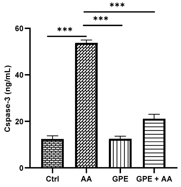
Background/Objectives: Ulcerative colitis (UC) is a chronic, relapsing inflammatory bowel disease (IBD) that poses a significant gastroenterological challenge. Methods: This study investigates the protective effects of garlic peel extract (GPE) in a rat model of acetic acid (AA)-induced colitis. Rats received oral GPE (100 mg/kg) for 14 days prior to AA administration, and this continued for 14 days post-induction. Results: GC-MS analysis of GPE identified several key phytochemicals, primarily methyl esters of fatty acids (62.47%), fatty acids (10.36%), fatty acid derivatives (6.75%), and vitamins (4.86%) as the major constituents. Other notable compounds included steroids, natural alcohols, organosulfur compounds, fatty aldehydes, carotenoids, sugars, and glucosinolates. GPE treatment significantly improved body weight and colon length. Biochemical analysis showed that GPE downregulated the levels of the pro-inflammatory cytokines interleukin-1 (IL-1), IL-6, IL-17, tumor necrosis factor-alpha (TNF-α), and nuclear factor-kappa B (NF-κB), compared to the colitis (AA) group. Additionally, GPE reduced the oxidative stress (OS) biomarkers, including myeloperoxidase (MPO) and malondialdehyde (MDA), as well as caspase-3, a marker for apoptosis. Furthermore, GPE treatment resulted in enhanced activities of the enzymatic antioxidants catalase (CAT) and superoxide dismutase (SOD), along with increased levels of the anti-inflammatory cytokine IL-10. These findings were supported by histological evidence. Conclusions: Collectively, GPE holds promise as a therapeutic strategy for UC, owing to its natural bioactive compounds and their potential synergistic anti-inflammatory, antioxidant, and anti-apoptotic effects.

Keywords: GC-MS; anti-inflammatory; antioxidants; cytokines; fatty acid esters; garlic peel extract; natural therapy; ulcerative colitis.


Conflict of interest statement

The authors declare no conflicts of interest.

Figures

Figure 1
Figure 1
GC-MS total ion chromatogram (TIC) of analyzed GPE.
Figure 2
Figure 2
Effect of GPE on final body weight (A) and colon length (B) in AA-induced UC rats. Data expressed as mean values ± SD (n = 6/group). ** = p < 0.001 and *** = p < 0.0001 vs. the AA group. Representative pictures of colon length (C) in all experimental groups.
Figure 3
Figure 3
Effect of GPE on pro- and anti-inflammatory mediators (A) IL-1, (B) IL-6, (C) IL-17, (D) TNF-α, (E) IL-10, and (F) the transcription factor NF-κB in AA-induced UC rats. Data expressed as mean values ± SD (n = 6/group). ** = p < 0.001 and *** = p < 0.0001 vs. the AA group.
Figure 4
Figure 4
Effect of GPE on oxidative stress biomarkers (A) CAT, (B) SOD, (C) MDA, and (D) MPO in AA-induced UC rats. Data expressed as mean values ± SD (n = 6/group). *** = p < 0.0001 vs. the AA group.
Figure 5
Figure 5
Effect of GPE on levels of caspase-3 in AA-induced UC rats. Data expressed as mean values ± SD (n = 6/group). *** = p < 0.0001 vs. the AA group.
Figure 6
Figure 6
Photomicrograph of histologically analyzed H&E-stained colon sections in all experimental groups: (A) control rats (Ctrl group) showing normal mucosal structure with intact epithelial surface (M), muscular layer (Mm), and submucosa (Sm); (B) rats with AA-induced colitis (AA group) showing histopathological alterations evidenced by severe mucosal damage, loss of goblet cells (arrowheads), and necrosis of mucosa (M) associated with marked infiltration of mononuclear cells (arrow); (C) GPE-treated rats (GPE group) showing almost nearly normal mucosal structure (M), muscular layer (Mm), and submucosa (Sm); (D) GPE-treated rats with colitis (GPE + AA group) showing improvement in histological structure with slight loss of some surface columnar epithelial cells (arrowhead) and minimal necrosis of mucosa (M). Original magnification: 100×. (E) Scoring of histology sections. Data expressed as mean ± SD (n = 6/group). *** = p < 0.0001 vs. AA group.

Similar articles

References

    1. Dakhli N., Rtibi K., Arrari F., Ayari A., Sebai H. Prophylactic coloprotective effect of Urtica dioica leaves against dextran sulfate sodium (DSS)-induced ulcerative colitis in rats. Medicina. 2023;59:1990. doi: 10.3390/medicina59111990. - DOI - PMC - PubMed
    1. Li S., Zhuge A., Chen H., Han S., Shen J., Wang K., Xia J., Xia H., Jiang S., Wu Y., et al. Sedanolide alleviates DSS-induced colitis by modulating the intestinal FXR-SMPD3 pathway in mice. J. Adv. Res. 2025;69:413–426. doi: 10.1016/j.jare.2024.03.026. - DOI - PMC - PubMed
    1. Feng J., Liu Y., Zhang C., Ji M., Li C. Phytic acid regulates proliferation of colorectal cancer cells by downregulating NF-kB and β-catenin signalling. Eur. J. Inflamm. 2023;21:1–14. doi: 10.1177/1721727X231182622. - DOI
    1. Alfwuaires M.A., Algefare A.I., Afkar E., Salam S.A., El-Moaty H.I.A., Badr G.M. Immunomodulatory assessment of Portulaca oleracea L. extract in a mouse model of colitis. Biomed. Pharmacother. 2021;143:112148. doi: 10.1016/j.biopha.2021.112148. - DOI - PubMed
    1. El Mahdy R.N., Nader M.A., Helal M.G., Abu-Risha S.E., Abdelmageed M.E. Eicosapentaenoic acid mitigates ulcerative colitis-induced by acetic acid through modulation of NF-κB and TGF-β/EGFR signaling pathways. Life Sci. 2023;327:121820. doi: 10.1016/j.lfs.2023.121820. - DOI - PubMed

LinkOut - more resources