. 2025 Jul 17;18(7):1050.
doi: 10.3390/ph18071050.

Bone Marrow Mesenchymal Stem Cell-Derived Exosomes Modulate Chemoradiotherapy Response in Cervical Cancer Spheroids

Affiliations

Bone Marrow Mesenchymal Stem Cell-Derived Exosomes Modulate Chemoradiotherapy Response in Cervical Cancer Spheroids

Kesara Nittayaboon et al. Pharmaceuticals (Basel). .

Abstract

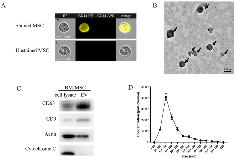
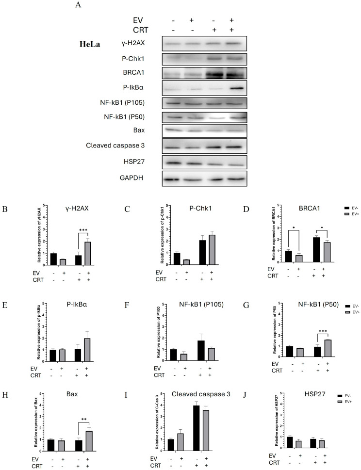
Background: Bone marrow mesenchymal stem cells (BM-MSCs) are significant in chemo- and radiotherapy resistance. Previous research has focused on BM-MSCs, demonstrating their functional involvement in cancer progression as mediators in the tumor microenvironment. They play multiple roles in tumorigenesis, angiogenesis, and metastasis. BM-MSC-derived exosomes (BM-MSCs-exo) are small vesicles, typically 50-300 nm in diameter, isolated from BM-MSCs. Some studies have demonstrated the tumor-suppressive effects of BM-MSCs-exo. Objective: This study aimed to investigate their role in modulating the impact of chemoradiotherapy (CRT) in different types of cervical cancer spheroid cells. Methods: The spheroids after treatment were subject to size measurement, cell viability, and caspase activity. Then, the molecular mechanism was elucidated by Western blot analysis. Results: We observed a reduction in spheroid size and an increase in cell death in HeLa spheroids, while no significant changes in size or cell viability were found in SiHa spheroids. At the molecular level, CRT treatment combined with BM-MSCs-exo in HeLa spheroids induced apoptosis through the activation of the NF-κB pathway, specifically via the NF-κB1 (P50) transcription factor, leading to the upregulation of apoptosis-related molecules. In contrast, CRT combined with BM-MSCs-exo in SiHa spheroids exhibited an opposing effect: although cellular viability decreased, caspase activity also decreased, which correlated with increased HSP27 expression and the subsequent downregulation of apoptotic molecules. Conclusion: Our study provides deeper insight into the potential of BM-MSCs-exo in cervical cancer treatment, supporting the development of more effective and safer therapeutic strategies for clinical application.

Keywords: bone marrow mesenchymal stem cells (BM-MSCs); cancer therapy; cervical cancer; chemo- and radiotherapy resistance; exosomes.


Conflict of interest statement

The authors declare no conflicts of interest.

Figures

Figure 1
Figure 1
Research workflow. This flowchart represents the experimental design. BM-MSCs-exo were isolated and characterized (A,B). Cervical spheroid cell cultures (C) were pre-treated (D) using BM-MSCs-exo, and then, CRT treatment was performed (E). After all treatment, the spheroids were incubated for 72 h, and functional analysis was then conducted (F). BM-MSC: Bone marrow mesenchymal stem cells; BM-MSC-exo: Bone marrow mesenchymal stem cell-derived exosome; Exo: Exosome; TEM: Transmission electron microscope; CRT: Chemoradiotherapy; Cis: Cisplatin; Gy: Gray. Created in BioRender. Nittayaboon, K. (2025).
Figure 2
Figure 2
Characterization of BM-MSCs-exo (A). CD44-positive and CD14-negative cell surface marker staining indicated the presence of BM-MSCs (B). The transmission electron microscope (TEM) showed cup-shape morphology of the BM-MSCs-exo indicated by the black arrows (C). Western blot analysis showed the protein expression of EV markers, including CD63 and CD9. Actin was used as housekeeping protein, and Cytochrome C served to control EV purity (D). The size distribution of BM-MSCs-exo was analyzed using nanoparticle tracking analysis (NTA). BF: Bright field; CD44-PE; CD44 protein conjugated with the fluorescent molecule phycoerythrin; CD14-APC: CD14 protein conjugated with the fluorescent molecule allophycocyanin; BM-MSC-exo: Bone marrow mesenchymal stem cell-derived exosome; MSC: Mesenchymal stem cells; EV: Extracellular vesicle; BF: Bright field; CD44-PE; CD44 protein conjugated with the fluorescent molecule phycoerythrin; CD14-APC: CD14 protein conjugated with the fluorescent molecule allophycocyanin.
Figure 3
Figure 3
The effect of BM-MSCs-exo combined with CRT treatment on cervical cancer spheroids. CRT treatment resulted in a slight decrease in spheroid size in HeLa but not in SiHa spheroids (A). The morphology of HeLa (upper panel) and SiHa (lower panel) spheroids was captured using inverted microscopy at 10× magnification (scale bar = 500 µm). (B,C) The sizes of SiHa and HeLa spheroids were measured using ImageJ software. Three independent experiments were conducted. Student’s t-test was used to determine statistical significance (** p < 0.01). EV: Extracellular vesicle; CRT: Chemoradiotherapy.
Figure 4
Figure 4
Spheroid viability and caspase activity were assessed using the LIVE/DEAD® Cell Imaging Kit and ApoLive-Glo™ Multiplex Assay (A). The upper panel shows the cell morphology and fluorescent signals of live cells (green fluorescence) and dead cells (red fluorescence). The lower panel presents the percentage of cell viability and caspase activity in cervical cancer spheroids, (B) HeLa and (C) SiHa. The images were captured using an inverted microscope and a LionheartFX live cell imager at 4× magnification (scale bar = 500 µm). The results are presented as the mean ± SD from three independent experiments. Student’s t-test was used to determine statistical significance (* p < 0.05; ** p < 0.01; **** p < 0.0001). EV: Extracellular vesicle; CRT: Chemoradiotherapy.
Figure 5
Figure 5
Molecular mechanisms by which BM-MSCs-exo enhanced the effect of CRT treatment on HeLa spheroids through the activation of apoptotic pathways (A). The expression levels of proteins involved in DNA damage, NF-κB signaling, and apoptosis pathways. The relative expression of each protein was normalized to GAPDH. Quantified proteins of interest included the following: γ-H2AX (B), p-Chk1 (C), p-BRCA1 (D), p-IκBα (E), NF-κB1 (P100) (F), NF-κB1 (P50) (G), Bax (H), cleaved caspase-3 (I), and HSP27 (J). Data are presented as the mean ± SD from three independent experiments. Student’s t-test was used to determine statistical significance (* p < 0.05; ** p < 0.01; *** p < 0.001). EV: Extracellular vesicle; CRT: Chemoradiotherapy.
Figure 6
Figure 6
Molecular mechanisms by which BM-MSCs-exo enhanced CRT resistance in SiHa spheroids through the downregulation of NF-κB and apoptotic molecules (A). The expression levels of proteins involved in DNA damage, NF-κB signaling, and apoptosis pathways. The relative expression of each protein was normalized to GAPDH. Quantified proteins of interest included γ-H2AX (B), p-Chk1 (C), p-BRCA1 (D), p-IκBα (E), NF-κB1 (P100) (F), NF-κB1 (P50) (G), Bax (H), cleaved caspase-3 (I), and HSP27 (J), with all intensities normalized to GAPDH band intensity. Data are presented as the mean ± SD from three independent experiments. Student’s t-test was used to assess statistical significance (* p < 0.05; ** p < 0.01; **** p < 0.0001). EV: Extracellular vesicle; CRT: Chemoradiotherapy.
Figure 7
Figure 7
Proposed molecular mechanism of BM-MSC-derived exosomes (BM-MSCs-exo), illustrating their dual role in promoting apoptosis and contributing to chemoradiotherapy (CRT) resistance in cervical cancer spheroids. The diagram highlights NF-κB activation and its downstream signaling pathways as potential mediators of these effects. Experimentally validated mechanisms are indicated by solid arrows, whereas hypothetical or inferred interactions are represented by dashed arrows. Figure created with BioRender. Nittayaboon, K. (2025).

Similar articles

References

    1. Hmadcha A., Martin-Montalvo A., Gauthier B.R., Soria B., Capilla-Gonzalez V. Therapeutic potential of mesenchymal stem cells for cancer therapy. Front. Bioeng. Biotechnol. 2020;8:43. doi: 10.3389/fbioe.2020.00043. - DOI - PMC - PubMed
    1. Papaccio F., Paino F., Regad T., Papaccio G., Desiderio V., Tirino V. Concise review: Cancer cells, cancer stem cells, and mesenchymal stem cells: Influence in cancer development. Stem Cells Transl. Med. 2017;6:2115–2125. doi: 10.1002/sctm.17-0138. - DOI - PMC - PubMed
    1. Ji N., Yu J.-W., Ni X.-C., Wu J.-G., Wang S.-L., Jiang B.-J. Bone marrow-derived mesenchymal stem cells increase drug resistance in CD133-expressing gastric cancer cells by regulating the PI3K/AKT pathway. Tumor Biol. 2016;37:14637–14651. doi: 10.1007/s13277-016-5319-0. - DOI - PubMed
    1. Zhang X., Tu H., Yang Y., Fang L., Wu Q., Li J. Mesenchymal stem cell-derived extracellular vesicles: Roles in tumor growth, progression, and drug resistance. Stem Cells Int. 2017;2017:1758139. doi: 10.1155/2017/1758139. - DOI - PMC - PubMed
    1. Ji R., Zhang B., Zhang X., Xue J., Yuan X., Yan Y., Wang M., Zhu W., Qian H., Xu W. Exosomes derived from human mesenchymal stem cells confer drug resistance in gastric cancer. Cell Cycle. 2015;14:2473–2483. doi: 10.1080/15384101.2015.1005530. - DOI - PMC - PubMed

LinkOut - more resources