. 2025 Jul 4;17(7):879.
doi: 10.3390/pharmaceutics17070879.

Crotoxin-Loaded Silica Nanoparticles: A Nanovenom Approach

Affiliations

Crotoxin-Loaded Silica Nanoparticles: A Nanovenom Approach

Florencia Silvina Conti et al. Pharmaceutics. .

Abstract

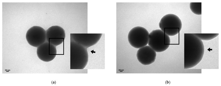
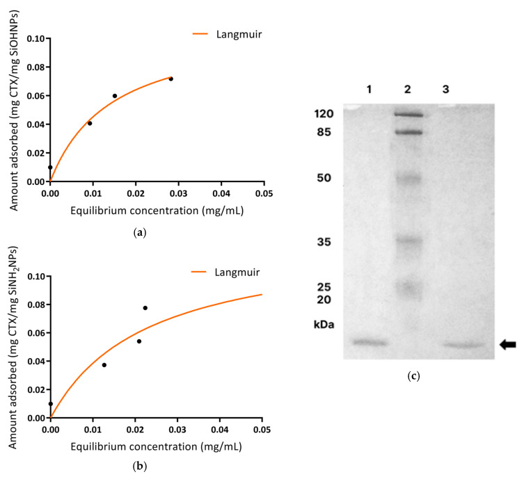
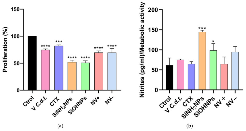
Background: Ophidism is a globally neglected health problem. In Argentina, Crotalus durissus terrificus (C.d.t., South American rattlesnake) is one of the species of greatest medical importance since its venom contains mainly crotoxin (CTX), a potent enzyme-toxin with PLA2 activity, which is responsible for its high lethality. Objective: In this work, we aimed to generate nanovenoms (NVs), complexes formed by CTX adsorbed onto 150 nm silica nanoparticles (SiNPs), and to study their physicochemical, biological, and immunomodulatory activities for potential use as adjuvants (ADJs) in antivenom (AV) production. Methods: CTX was isolated and corroborated by SDS-PAGE. Then, CTX was adsorbed on the synthetized Stöber SiNPs' surfaces, forming a monolayer and retaining its biological activity (as observed by the MTT cell proliferation assay using the THP-1 cell line). Results: Immunomodulatory activity revealed a high pro-inflammatory (IL-1β) response induced by SiNPs followed by NVs. In the case of the anti-inflammatory response, NVs presented significant differences for TGF-β only after cell activation with LPS. No significant differences were observed in IL-10 levels. Conclusions: Thus, these results suggest that NVs together with SiNPs could increase immunogenicity and enhance immune response, turning them into potential tools for the generation of new antivenoms.

Keywords: PLA2; SiNPs; adjuvants; nanovenoms; serotherapy; snake venom.


Conflict of interest statement

The authors declare no conflicts of interest.

Figures

Figure 1
Figure 1
Characterization of SiNPs. (a). Dynamic light scattering (DLS). Histogram of the diameters of the SiNPs obtained by DLS. Transmission Electron Microscopy (TEM) micrographs. Bare silica morphologies of SiOHNPs (b) and SiNH2NPs (c) obtained according to the Stöber method. Scale bar: 100 nm. (d). FTIR: infrared spectra of SiOHNPs (green) and SiNH2NPs (orange).
Figure 1
Figure 1
Characterization of SiNPs. (a). Dynamic light scattering (DLS). Histogram of the diameters of the SiNPs obtained by DLS. Transmission Electron Microscopy (TEM) micrographs. Bare silica morphologies of SiOHNPs (b) and SiNH2NPs (c) obtained according to the Stöber method. Scale bar: 100 nm. (d). FTIR: infrared spectra of SiOHNPs (green) and SiNH2NPs (orange).
Figure 2
Figure 2
Isolation and quantification of purified CTX. (a). Elution plot. Elution chromatographic profile resulting from FPLC. The elution peaks refer to convulxin (1), crotoxin (2), and crotamine (3). (b). SDS-Page: polyacrylamide gel electrophoresis (14%) of whole C.d.t. venom and CTX. Molecular mass marker (1), C.d.t. venom (2), and isolated CTX (3). Black arrows indicate the venom proteins: CVX (A), LAAO (B), CTX (C), and CTM (D).
Figure 3
Figure 3
Desorption of CTX. Stability over time of CTX adsorbed onto NVs+/−. The graph shows the percentage of desorption (mean ± SD) of this protein in both types of NVs at different times (30 min, 1 and 2 h).
Figure 4
Figure 4
TEM micrograph. Micrographs of NV− (a) and NV+ (b) at 150 nm. The adsorbed CTX is indicated by a black arrow. Scale bar: 20 nm. (c). FTIR: infrared spectra of NV− (green) and NV+ (orange). The absorption bands, indicated with black arrows, of disulfide bridge bonds at 425 cm−1 and amine groups of proteins at 1600 cm−1 and 3400 cm−1 can be observed.
Figure 4
Figure 4
TEM micrograph. Micrographs of NV− (a) and NV+ (b) at 150 nm. The adsorbed CTX is indicated by a black arrow. Scale bar: 20 nm. (c). FTIR: infrared spectra of NV− (green) and NV+ (orange). The absorption bands, indicated with black arrows, of disulfide bridge bonds at 425 cm−1 and amine groups of proteins at 1600 cm−1 and 3400 cm−1 can be observed.
Figure 5
Figure 5
Adsorption isotherms. Isotherms of NV− (a) and NV+ (b) with different CTX concentrations (0.010, 0.050, 0.075, 0.1, 0.2). Both graphics adjust to the Langmuir model. Maximum adsorption capacity = 0.110 mg/mg SiOHNPs, R2 = 0.9452, and Km = 0.0143 mg/mL (a); maximum adsorption capacity = 0.127 mg/mg SiNH2NPs, R2 = 0.9142, and Km = 0.0229 mg/mL (b). (c). SDS-PAGE: bands corresponding to CTX desorbed from NV+ (1) and NV− (3) can be observed. The black arrow indicates CTX, and the second column (2) corresponds to the molecular marker.
Figure 6
Figure 6
(a) Cell proliferation assay (MTT): cell proliferation (%) assay using THP-1 cell line. (b). Nitrites: nitrite levels in the SN of the different treatments with respect to the metabolic activity. Asterisks indicate significant differences between treatments (* p > 0.05;; *** p > 0.001; **** p > 0.0001, n = 3).
Figure 7
Figure 7
(a) IL-1β: ELISA of the cell SN of the different treatments. (b). TGF-β: ELISA of the cell SN after the LPS activation of the different treatments. Asterisks indicate significant differences between treatments (* p > 0.05; ** p > 0.01; *** p > 0.001, n = 3).

Similar articles

References

    1. World Health Organization Guidelines for the Production Control and Regulation of Snake Antivenom Immunoglobulins. 2016. [(accessed on 29 June 2025)]. Available online: https://extranet.who.int/prequal/vaccines/guidelines-production-control-....
    1. Amaral C.F., Magalhaes R.A., de Renzende N.A. Respiratory involvement secondary to crotalidophidian bite (Crotalus durissus) Rev. Inst. Med. Trop. São Paulo. 1991;33:251–255. doi: 10.1590/S0036-46651991000400002. - DOI - PubMed
    1. Baranauskas V., Dourado D.M., Jingguo Z., da Cruz-Höfling M.A. Characterization of the Crotalus durissus terrificus venom by atomic force microscopy. J. Vac. Sci. Technol. B Microelectron. Nanometer Struct. Process. Meas. Phenom. 2002;20:1317–1320. doi: 10.1116/1.1486007. - DOI
    1. Georgieva D., Ohler M., Seifert J., von Bergen M., Arni R.K., Genov N., Betzel C. Snake venomic of Crotalus durissus terrificus correlation with pharmacological activities. J. Proteome Res. 2010;9:2302–2316. doi: 10.1021/pr901042p. - DOI - PubMed
    1. Chippaux J.P. Snake-bites: Appraisal of the global situation. Bull. World Health Organ. 1998;76:515–524. - PMC - PubMed

LinkOut - more resources