. 2025 Jul 14;17(7):910.
doi: 10.3390/pharmaceutics17070910.

Application of Standardized Rosa damascena Stem Cell-Derived Exosomes in Dermatological Wound Healing and Scar Management: A Retrospective Case-Series Study with Long-Term Outcome Assessment

Affiliations

Application of Standardized Rosa damascena Stem Cell-Derived Exosomes in Dermatological Wound Healing and Scar Management: A Retrospective Case-Series Study with Long-Term Outcome Assessment

Lidia Majewska et al. Pharmaceutics. .

Abstract

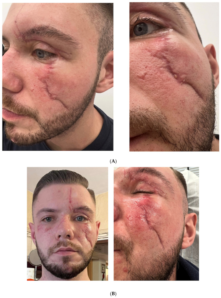
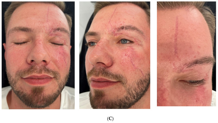
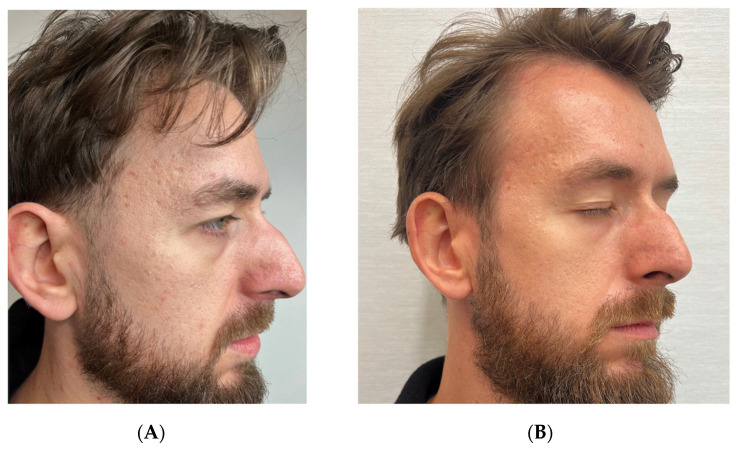
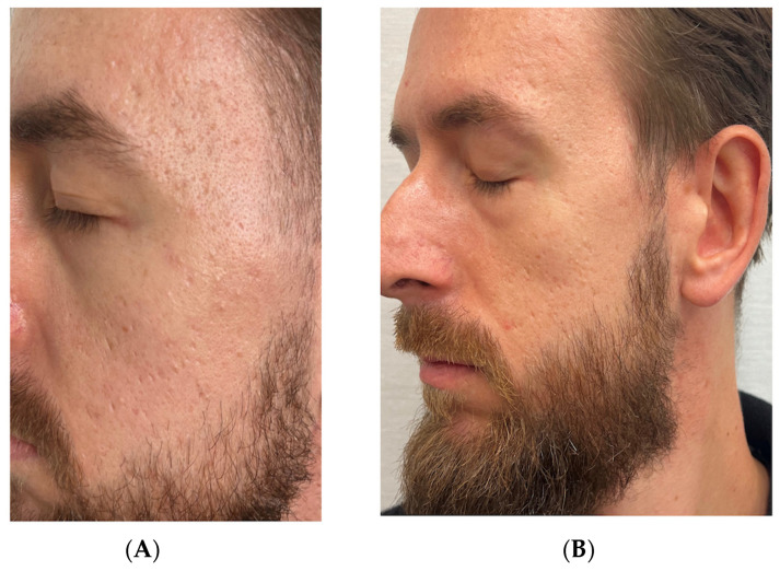
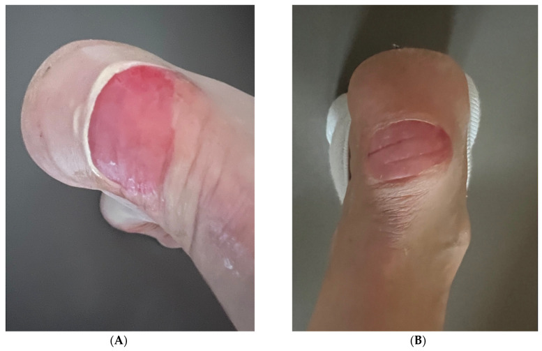
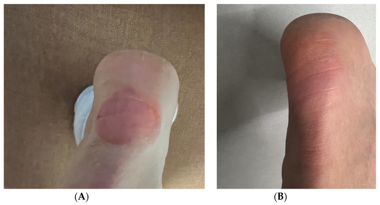
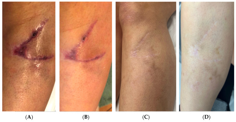
Background: Scar formation and impaired wound healing represent significant challenges in dermatology and aesthetic medicine, with limited effective treatment options currently available. Objectives: To evaluate the efficacy and long-term outcomes of Damask rose stem-cell-derived exosome (RSCE) therapy in the management of diverse dermatological conditions, including traumatic wounds, surgical scars, and atrophic acne scars. Methods: We conducted a case series study from June 2023 to November 2024, documenting four cases with different types of skin damage treated with lyophilized RSCE products. Treatment protocols included a variety of delivery methods such as topical application, microneedling, and post-procedure care. Follow-up assessments were performed at intervals ranging from 7 days to 10 months. Results: All patients demonstrated significant improvements in scar appearance, skin elasticity, hydration, and overall tissue quality. In traumatic facial injury, RSCE therapy facilitated reduction in scar contracture and improved functional outcomes. For atrophic acne scars, comparative treatment of facial sides showed enhanced results with RSCE addition. Acute wounds exhibited accelerated healing with reduced inflammation, while chronic wounds demonstrated improved epithelialization and long-term scar quality. Conclusions: This case series provides preliminary evidence suggesting that RSCE therapy may offer significant benefits in wound healing and scar management. The observed improvements in tissue regeneration, inflammatory modulation, and long-term aesthetic outcomes warrant further investigation through controlled clinical trials.

Keywords: atrophic acne scars; damask rose stem cells; exosome therapy; exosomes; extracellular vesicles; plant-derived exosome-like nanovesicles; post-surgical scars; regenerative dermatology; rose stem cell-derived exosomes; scar remodeling; skin regeneration; wound healing.


Conflict of interest statement

The authors declare no conflicts of interest.

Figures

Figure 1
Figure 1
Traumatic facial injury-treatment progression in a 24-year-old male patient. (A) Baseline appearance, showing severe facial scarring with contracture and hyperesthesia four months post-accident. (B) Clinical appearance at 30 days post-treatment initiation, showing initial improvement in scar depth and tissue mobility. (C) Final outcome at 110 days, demonstrating significant reduction in scar adhesion, improved eye closure functionality, and enhanced skin texture and elasticity.
Figure 1
Figure 1
Traumatic facial injury-treatment progression in a 24-year-old male patient. (A) Baseline appearance, showing severe facial scarring with contracture and hyperesthesia four months post-accident. (B) Clinical appearance at 30 days post-treatment initiation, showing initial improvement in scar depth and tissue mobility. (C) Final outcome at 110 days, demonstrating significant reduction in scar adhesion, improved eye closure functionality, and enhanced skin texture and elasticity.
Figure 2
Figure 2
Atrophic acne scar-treatment progression on the right side of face (RSCE-enhanced treatment) in a 32-year-old male patient. (A) Baseline appearance, showing Grade 4 (severe) atrophic acne scarring with prominent boxcar and rolling scars. (B) Ten-month follow-up, demonstrating significant improvement to Grade 2, with markedly reduced scar depth, enhanced skin texture, and improved surface regularity.
Figure 3
Figure 3
Atrophic acne scar-treatment progression on the left side of face (standard treatment) in a 32-year-old male patient. (A) Baseline appearance, showing Grade 3 (moderate to severe) atrophic acne scarring with multiple shallow-to-moderate-depth scars. (B) Ten-month follow-up after standard treatment protocol (subcision, hyaluronic acid, and microneedling), showing improvement to Grade 2, with moderate reduction in scar visibility and improved skin texture.
Figure 4
Figure 4
Acute heel wound-healing progression in a 51-year-old female patient during initial RSCE treatment phase. (A) Baseline presentation at 12 h post-injury, showing 2.5 cm diameter acute wound with exposed dermis, active bleeding, and significant periwound erythema and inflammation. (B) Twenty-four hours after RSCE treatment initiation, demonstrating marked reduction in periwound inflammation, decreased erythema, early epithelial migration, and absence of active bleeding with initial wound-healing response.
Figure 5
Figure 5
Final stages of acute heel wound-healing progression. (A) Clinical presentation at 72 h (day 3), showing advanced epithelialization with substantial wound-area reduction and continued resolution of periwound inflammation. (B) Day 7, final assessment, demonstrating complete wound closure with optimal healing outcome—full re-epithelialization, normal skin texture and elasticity, and absence of visible scarring or pigmentation abnormalities.
Figure 6
Figure 6
Lacerated leg wound-healing progression in a 42-year-old female patient treated with RSCE therapy. (A) Baseline presentation at 2 weeks post-injury, showing 4 cm oblique laceration with delayed healing, persistent erythema, and incomplete epithelialization despite standard care with Cicaplast Baume. (B) Day 18 post-injury (4 days after RSCE therapy initiation), demonstrating significant reduction in erythema and inflammation with approximately 80% wound closure and healthy epithelial advancement. (C) Six-month follow-up, revealing substantial reduction in scar prominence, with improved skin texture, minimal discoloration compared to surrounding skin, and excellent tissue integration. (D) Eight-month final assessment, showing near-complete integration with surrounding skin, demonstrating optimal coloration match, excellent pliability, and minimal scar visibility under casual observation.

Similar articles

  • Interventions for acne scars.
    Abdel Hay R, Shalaby K, Zaher H, Hafez V, Chi CC, Dimitri S, Nabhan AF, Layton AM. Abdel Hay R, et al. Cochrane Database Syst Rev. 2016 Apr 3;4(4):CD011946. doi: 10.1002/14651858.CD011946.pub2. Cochrane Database Syst Rev. 2016. PMID: 27038134 Free PMC article.
  • Laser therapy for treating hypertrophic and keloid scars.
    Leszczynski R, da Silva CA, Pinto ACPN, Kuczynski U, da Silva EM. Leszczynski R, et al. Cochrane Database Syst Rev. 2022 Sep 26;9(9):CD011642. doi: 10.1002/14651858.CD011642.pub2. Cochrane Database Syst Rev. 2022. PMID: 36161591 Free PMC article.
  • Pressure-garment therapy for preventing hypertrophic scarring after burn injury.
    Harris IM, Lee KC, Deeks JJ, Moore DJ, Moiemen NS, Dretzke J. Harris IM, et al. Cochrane Database Syst Rev. 2024 Jan 8;1(1):CD013530. doi: 10.1002/14651858.CD013530.pub2. Cochrane Database Syst Rev. 2024. PMID: 38189494 Free PMC article.
  • Negative pressure wound therapy for open traumatic wounds.
    Iheozor-Ejiofor Z, Newton K, Dumville JC, Costa ML, Norman G, Bruce J. Iheozor-Ejiofor Z, et al. Cochrane Database Syst Rev. 2018 Jul 3;7(7):CD012522. doi: 10.1002/14651858.CD012522.pub2. Cochrane Database Syst Rev. 2018. PMID: 29969521 Free PMC article.
  • Silicone gel sheeting for treating keloid scars.
    Tian F, Jiang Q, Chen J, Liu Z. Tian F, et al. Cochrane Database Syst Rev. 2023 Jan 3;1(1):CD013878. doi: 10.1002/14651858.CD013878.pub2. Cochrane Database Syst Rev. 2023. PMID: 36594476 Free PMC article.

References

    1. desJardins-Park H.E., Gurtner G.C., Wan D.C., Longaker M.T. From Chronic Wounds to Scarring: The Growing Health Care Burden of Under- and Over-Healing Wounds. Adv. Wound Care. 2022;11:496–510. doi: 10.1089/wound.2021.0039. - DOI - PMC - PubMed
    1. Meretsky C.R., Polychronis A., Schiuma A.T. A Comparative Analysis of the Advances in Scar Reduction: Techniques, Technologies, and Efficacy in Plastic Surgery. Cureus. 2024;16:e66806. doi: 10.7759/cureus.66806. - DOI - PMC - PubMed
    1. Han Y., Qin X., Lin W., Wang C., Yin X., Wu J., Chen Y., Chen X., Chen T. Microneedle-Based Approaches for Skin Disease Treatment. Nanomicro Lett. 2025;17:132. doi: 10.1007/s40820-025-01662-y. - DOI - PMC - PubMed
    1. Yang G., Waheed S., Wang C., Shekh M., Li Z., Wu J. Exosomes and Their Bioengineering Strategies in the Cutaneous Wound Healing and Related Complications: Current Knowledge and Future Perspectives. Int. J. Biol. Sci. 2023;19:1430–1454. doi: 10.7150/ijbs.80430. - DOI - PMC - PubMed
    1. Hao M., Duan M., Yang Z., Zhou H., Li S., Xiang J., Wu H., Liu H., Chang L., Wang D., et al. Engineered Stem Cell Exosomes for Oral and Maxillofacial Wound Healing. Front. Bioeng. Biotechnol. 2022;10:1038261. doi: 10.3389/fbioe.2022.1038261. - DOI - PMC - PubMed

LinkOut - more resources