. 2025 Jul 10;30(14):2923.
doi: 10.3390/molecules30142923.

Flipping the Target: Evaluating Natural LDHA Inhibitors for Selective LDHB Modulation

Affiliations

Flipping the Target: Evaluating Natural LDHA Inhibitors for Selective LDHB Modulation

Amanda El Khoury et al. Molecules. .

Abstract

Lactate dehydrogenase (LDH) catalyzes the reversible interconversion of pyruvate and lactate, coupled with the redox cycling of NADH and NAD+. While LDHA has been extensively studied as a therapeutic target, particularly in cancer, due to its role in the Warburg effect, LDHB remains underexplored, despite its involvement in the metabolic reprogramming of specific cancer types, including breast and lung cancers. Most known LDH inhibitors are designed against the LDHA isoform and act competitively at the active site. In contrast, LDHB exhibits distinct kinetic properties, substrate preferences, and structural features, warranting isoform-specific screening strategies. In this study, 115 natural compounds previously reported as LDHA inhibitors were systematically evaluated for LDHB inhibition using an integrated in silico and in vitro approach. Virtual screening identified 16 lead phytochemicals, among which luteolin and quercetin exhibited uncompetitive inhibition of LDHB, as demonstrated by enzyme kinetic assays. These findings were strongly supported by molecular docking analyses, which revealed that both compounds bind at an allosteric site located at the dimer interface, closely resembling the binding mode of the established LDHB uncompetitive inhibitor AXKO-0046. In contrast, comparative docking against LDHA confirmed their active-site binding and competitive inhibition, underscoring their isoform-specific behavior. Our findings highlight the necessity of assay conditions tailored to LDHB's physiological role and demonstrate the application of a previously validated colorimetric assay for high-throughput screening. This work lays the foundation for the rational design of selective LDHB inhibitors from natural product libraries.

Keywords: allosteric inhibition; cancer metabolism; isoform selectivity; lactate dehydrogenase B; natural products.


Conflict of interest statement

The authors declare no conflicts of interest.

Figures

Figure 1
Figure 1
The top-scoring natural compounds identified through molecular docking as potential allosteric inhibitors of LDHB. Docking was conducted using the crystal structure of LDHB (PDB ID: 7DBJ). The chemical structures and corresponding docking scores (in kcal/mol) of the 16 compounds that achieved scores equal to or better than that of AXKO-0046 (−7.6 kcal/mol), a known uncompetitive inhibitor of LDHB, are illustrated.
Figure 2
Figure 2
The evaluation of the top candidate compounds for LDHB inhibition. The inhibitory activity of the selected compounds was assessed using a 96-well endpoint colorimetric assay, as described in the Section 4. (A) The inhibition of LDHB activity by the top four candidates identified from virtual screening, compared to the known LDHB inhibitor AXKO-0046. The horizontal bars indicate the mean values from three independent experiments. The dashed line indicates 50% inhibition. Dose–response curves show the inhibitory effect of AXKO-0046 (B), luteolin (C), and quercetin (D) on LDHB activity. The percentage of LDHB inhibition was determined by comparing the enzyme activity in the presence of each compound to that of a control without any of the inhibitors. IC50 values were calculated using nonlinear regression analysis. The molecular structures of the compounds are shown in each panel.
Figure 3
Figure 3
A kinetic analysis of LDHB inhibition by luteolin and quercetin. The inhibitory effects of luteolin (A,B) and quercetin (C,D) on LDHB activity were assessed using varying concentrations of sodium lactate (A,C) and NAD+ (B,D). Lineweaver–Burk plots were constructed using linear regression analysis from the kinetic data (see Table 2), with each curve corresponding to a different inhibitor concentration (0–100 μM).
Figure 4
Figure 4
The predicted binding of AXKO-0046, luteolin, and quercetin on LDHB. Structural superposition shows LDHB in complex with NADH (blue sticks) bound at the active sites of each monomer (chain A: orange; chain C: cyan). AXKO-0046 (purple), luteolin (red), and quercetin (green) are all localized at the allosteric site formed at the dimer interface, distal to the catalytic site. This binding mode is consistent with their experimentally determined inhibition behavior, supporting an uncompetitive mechanism of action.
Figure 5
Figure 5
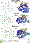
The molecular interactions of AXKO-0046 (A,B), luteolin (C,D), and quercetin (E,F) with the allosteric site of LDHB located in the interphase of chains A and C (in 7DBJ), illustrated as 2D interaction diagrams (left panels) and 3D hydrophobic surface maps (right panels). In the 2D diagrams, the amino acid residues from LDHB chains A and C involved in the interactions are labeled and highlighted with colored circles. These circles correspond to different interaction types, as indicated in the interaction key located in the bottom left panel of the figure. In the 3D hydrophobic surface maps, key hydrophobic (brown) and hydrophilic (blue) regions of the binding pocket are shown, along with hydrogen bonds and other non-covalent interactions. The color gradient reflects surface hydrophobicity, as explained in the key shown in the bottom right panel of the figure. The docking studies were carried out using PyRx v1.1 (AutoDock Vina). The 2D and 3D images were obtained using BIOVIA Discovery Studio.
Figure 6
Figure 6
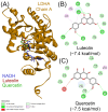
The docking of luteolin and quercetin with human LDHA. (A) The overall structure of LDHA (PDB ID: 4ZVV; chain A), showing the binding orientations of NADH (blue), luteolin (red), and quercetin (green) within the active site. The molecular interactions of luteolin (B) and quercetin (C) with LDHA are illustrated as 2D interaction diagrams. In these diagrams, interacting amino acid residues from LDHA chain A are labeled and highlighted with colored circles, corresponding to the different interaction types indicated in the interaction key in the bottom left panel. In (B,C), the binding scores are indicated in parentheses as kcal/mol.
Figure 7
Figure 7
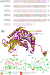
(A) The pairwise sequence alignment of human LDHA and LDHB. Identical amino acids are marked with an asterisk (*), conserved amino acids with a colon (:), and semi-conserved amino acids with a period (.). The residues are color-coded based on their chemical properties: acidic (red), basic (blue), polar (cyan), hydrophobic (green), and others (magenta). The alignment shows 75.1% identity (251 out of 334 residues) and 88.6% similarity (296 out of 334 residues), with notable substitutions near the active site and cofactor-binding regions. (B) Structural superposition of human LDHA (PDB ID: 4zvv; chain A, gold) bound to NADH (red) and LDHB (PDB ID: 1t2f; chain A, purple) bound to NAD+ (cyan), generated using UCSF ChimeraX v 1.9. Despite the overall structural conservation, differences are evident in the substrate-binding pockets and NADH/NAD+ interaction sites, highlighted in the two-dimensional interaction diagrams below the structure. Critical residues participating in hydrogen bonding and hydrophobic interactions are depicted, and ligand interaction types are labeled according to their interaction characteristics. These localized variations may influence isoform-specific ligand binding and inhibitory potency.
Figure 8
Figure 8
The key structural and functional differences between human LDHA and LDHB relevant to isoform-specific inhibitor design. Despite their high structural similarity, LDHA and LDHB differ in tissue distribution, substrate and cofactor preference, net charge, and inhibition mechanisms. LDHA is predominant in glycolytic tissues and catalyzes the conversion of pyruvate to lactate using NADH, while LDHB, expressed in oxidative tissues, favors the reverse reaction using NAD+. LDHB also shows substrate inhibition at high pyruvate levels. Importantly, LDHA is typically inhibited competitively at the active site, whereas LDHB is inhibited allosterically via uncompetitive mechanisms, as shown for luteolin and quercetin in this study. These differences highlight the need for isoform-specific assay conditions and targeted inhibitor development.

Similar articles

References

    1. Farhana A., Lappin S.L. StatPearls. StatPearls Publishing; Treasure Island, FL, USA: 2022. Biochemistry, lactate dehydrogenase. - PubMed
    1. Gladden L.B. Lactate metabolism: A new paradigm for the third millennium. J. Physiol. 2004;558:5–30. doi: 10.1113/jphysiol.2003.058701. - DOI - PMC - PubMed
    1. Adeva M., González-Lucán M., Seco M., Donapetry C. Enzymes involved in l-lactate metabolism in humans. Mitochondrion. 2013;13:615–629. doi: 10.1016/j.mito.2013.08.011. - DOI - PubMed
    1. Maeda M., Ko M., Mane M.M., Cohen I.J., Shindo M., Vemuri K., Serganova I., Blasberg R. Genetic and drug inhibition of LDH-A: Effects on murine gliomas. Cancers. 2022;14:2306. doi: 10.3390/cancers14092306. - DOI - PMC - PubMed
    1. Read J.A., Winter V.J., Eszes C.M., Sessions R.B., Brady R.L. Structural basis for altered activity of M- and H-isozyme forms of human lactate dehydrogenase. Proteins. 2001;43:175–185. doi: 10.1002/1097-0134(20010501)43:2<175::AID-PROT1029>3.0.CO;2-#. - DOI - PubMed

MeSH terms

LinkOut - more resources