. 2025 Jul 10;30(14):2925.
doi: 10.3390/molecules30142925.

From Antiretroviral to Antibacterial: Deep-Learning-Accelerated Repurposing and In Vitro Validation of Efavirenz Against Gram-Positive Bacteria

Affiliations

From Antiretroviral to Antibacterial: Deep-Learning-Accelerated Repurposing and In Vitro Validation of Efavirenz Against Gram-Positive Bacteria

Ezzeldin Saleh et al. Molecules. .

Abstract

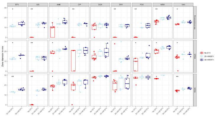
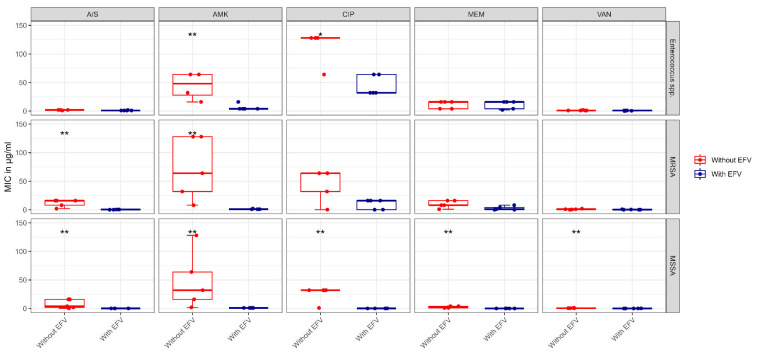
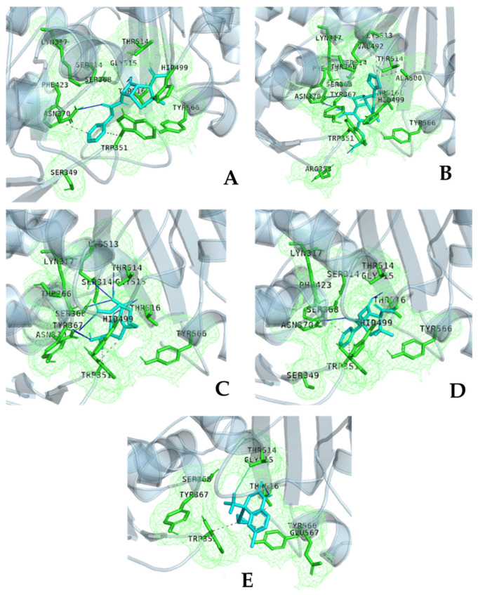
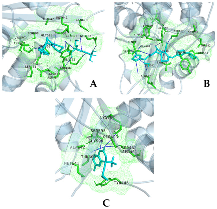
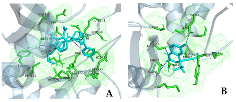
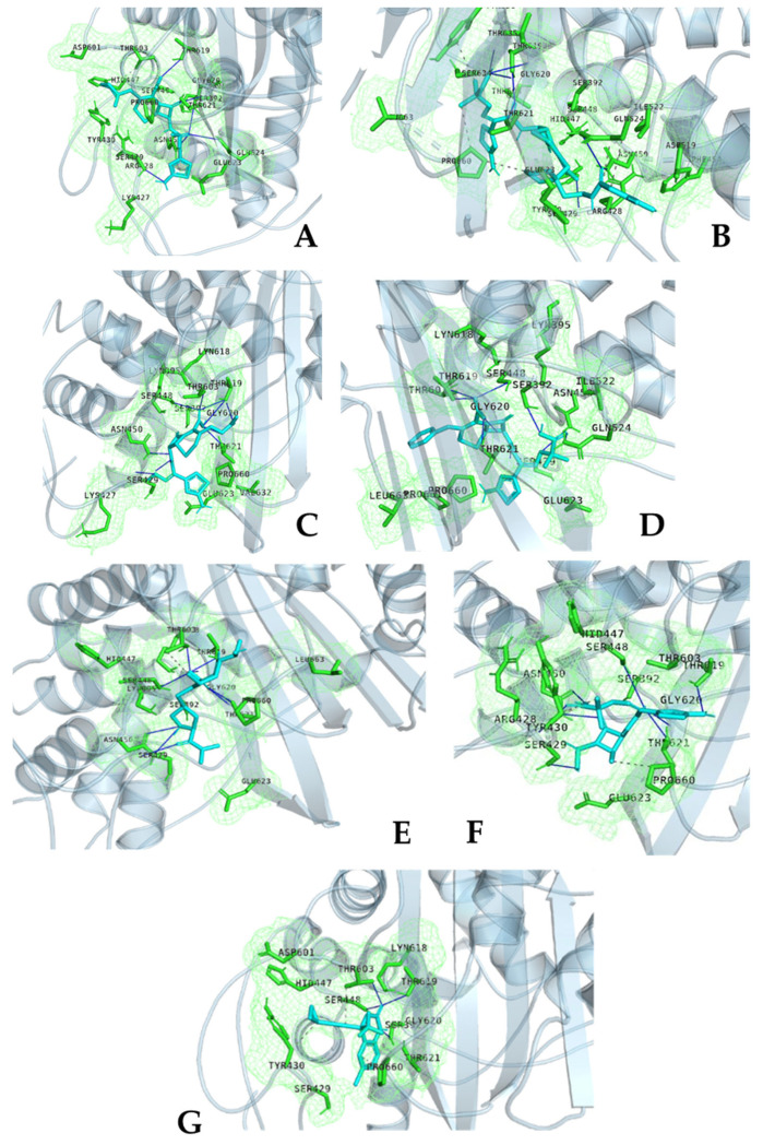
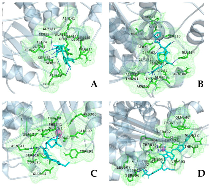
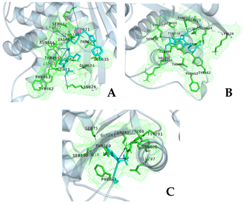
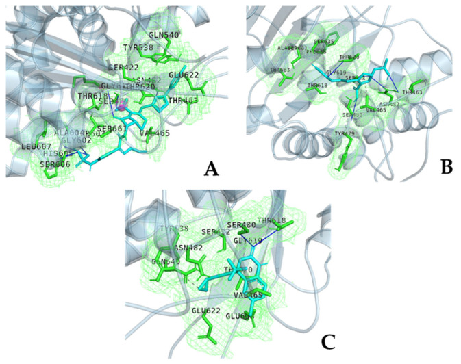
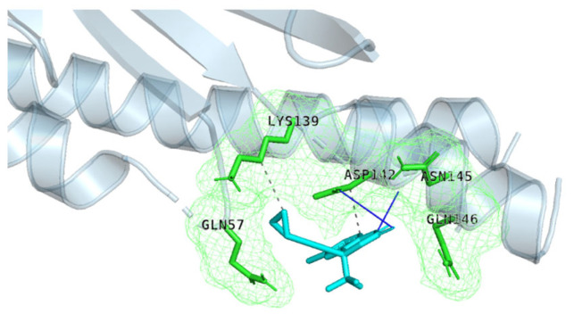
The repurposing potential of Efavirenz (EFV), a clinically established non-nucleoside reverse transcriptase inhibitor, was comprehensively evaluated for its in vitro antibacterial effect either alone or in combination with other antibacterial agents on several Gram-positive clinical strains showing different antibiotic resistance profiles. The binding potential assessed by an in silico study included Penicillin-binding proteins (PBPs) and WalK membrane kinase. Despite the relatively high minimum inhibitory concentration (MIC) limiting the use of EFV as a single antibacterial agent, it exhibits significant synergistic activity at sub-MIC levels when paired with various antibiotics against Enterococcus species and Staphylococcus aureus. EFV showed restored sensitivity of β-lactams against Methicillin-resistant S. aureus (MRSA). It increased the effectiveness of antibiotics tested against Methicillin-sensitive S. aureus (MSSA). It also helped to overcome the intrinsic resistance barrier for several antibiotics in Enterococcus spp. In silico binding studies aligned remarkably with experimental antimicrobial testing results and highlighted the potential of EFV to direct the engagement of PBPs with moderate to strong binding affinities (pKa 5.2-6.1). The dual-site PBP2 binding mechanism emerged as a novel inhibition strategy, potentially circumventing resistance mutations. Special attention should be paid to WalK binding predictions (pKa = 4.94), referring to the potential of EFV to interfere with essential regulatory pathways controlling cell wall metabolism and virulence factor expression. These findings, in general, suggest the possibility of EFV as a promising lead for the development of new antibacterial agents.

Keywords: Efavirenz; antibacterial activity; drug repurposing; in silico binding study.


Conflict of interest statement

The authors declare no conflicts of interest.

Figures

Scheme 1
Scheme 1
Chemical structure of the non-nucleoside reverse transcriptase inhibitor Efavirenz.
Figure 1
Figure 1
Effect of 25 mM and 50 mM of EFV on the diameter zone for eight antibiotics against Enterococcus spp., MRSA, and MSSA isolates. The asterisks indicate statistically significant change, * p value < 0.05, ** p value < 0.01. Disk diffusion assay showing the antibacterial effect of EFV (25 mM and 50 mM) by the zone of inhibition diameter (mm), alone or in combination with eight selected antibiotics against clinical isolates of Enterococcus spp. (n = 5), MRSA (n = 5), and MSSA (n = 5). EFV showed a significant dose-dependent antibacterial effect on the study isolates. EFV also restored sensitivity to antibiotics with no baseline activity. Significant increases in the inhibition zone with one or both concentrations were observed for β-lactams (Meropenem (MEM), Ampicillin/Sulbactam (A/S), Cefoxitine (FOX)) and aminoglycosides (Amikacin (AMK)) in MSSA and/or MRSA, while Enterococcus spp. showed a significant change with β-lactams, Vancomycin (VAN), and quinolones (Ciprofloxacin (CIP)) (p < 0.05, asterisks). Data represent median ± interquartile range (IQR); statistics: Kruskal–Wallis/Dunn’s test (multi-group) or Wilcoxon rank-sum test (two-group).
Figure 2
Figure 2
MIC of five antibiotics against Enterococcus spp., MRSA, and MSSA isolates without and with ½ × MIC EFV. The asterisks indicate statistically significant change, * p value < 0.05, ** p value < 0.01. MIC (μg/mL) of five antibiotics against Enterococcus spp. (n = 5), MRSA (n = 5), and MSSA (n = 5), tested alone (control) and with ½ × MIC EFV (8 μg/mL). EFV significantly reduced MICs for MSSA (all antibiotics (4–256-fold reduction; p < 0.05)), MRSA (AMK (64→1 μg/mL)), and A/S (16→0.5 μg/mL; p < 0.05). Enterococcus: AMK (64→4 μg/mL; p = 0.009) and CIP (128→32 μg/mL; p = 0.011). Asterisks denote statistical significance (Wilcoxon rank-sum test). Boxes: median (line), IQR (box), range (whiskers).
Figure 3
Figure 3
Comparative interaction fingerprints of β-lactam antibiotics and EFV within S. aureus PBP1. Panels (AD) illustrate the binding modes of different drugs (cyan) (A) amoxicillin, (B) cefuroxime, (C) imipenem, and (D) penicillin G within the catalytic pocket of PBP1, characterized by the covalent acylation of the catalytic Ser304 and conserved H-bonding with residues such as Asp267, Gln285, Lys300, and Asn308. These β-lactams also form a salt bridge between their carboxylate moieties and Lys544, stabilizing their transition-state mimicry. Panel (E) shows EFV binding in a distal allosteric site, approximately 15 Å from Ser304. EFV is stabilized via hydrophobic interactions with Trp351, Phe423, and Tyr566, H-bonds with Asn370 and Thr516, and a halogen bond between its fluorine atom and Thr514. Green meshes represent interaction fingerprints derived from docking pose analysis, highlighting key pharmacophoric features. While β-lactams uniformly engage the catalytic core, EFV adopts a non-canonical, allosteric binding mode that avoids direct interference with catalytic residues. This mechanistic divergence may underlie the ability of EFV to evade β-lactam resistance mechanisms and support its development as a novel antibacterial scaffold. Cyan represents the ligand (EFV or β-lactams), green meshes denote the pocket perimeter or interaction fingerprints, and grey depicts the protein backbone.
Figure 4
Figure 4
Comparative binding modes of Ceftaroline, Ceftobiprole, and EFV at the catalytic site of S. aureus PBP2a. The drug (cyan) (A) Ceftaroline engages the catalytic site with 11 H-bonds involving residues such as Thr600, Ser462, Glu602, and Lys597, mimicking the native D-Ala-D-Ala substrate. Hydrophobic interactions with Tyr446 and Thr582 further stabilize the complex, enabling the effective covalent acylation of the catalytic Ser400. (B) Ceftobiprole binds similarly but with a streamlined polar network (nine H-bonds), notably forming strong interactions with Ser400, Gln521, and Glu602. It lacks an interaction with Lys597 but compensates through compact hydrophobic engagement with Tyr446. (C) EFV adopts a distinct, non-catalytic binding pattern. It forms only four H-bonds (e.g., with Ser462, Ser598) and lacks contacts with Ser400 or Glu602. Instead, it stabilizes through hydrophobic contacts with Tyr446 and Ala642, occupying a peripheral sub-pocket. Green meshes highlight key interaction fingerprints. The divergence in polar and spatial engagement explains the high inhibitory potency of β-lactams versus the limited catalytic interference by EFV. Cyan represents the ligand (EFV or β-lactams), green meshes denote the pocket perimeter or interaction fingerprints, and grey depicts the protein backbone.
Figure 5
Figure 5
Divergent allosteric binding strategies of Ceftaroline and EFV within S. aureus PBP2a. The drug (cyan) (A) Ceftaroline anchors to the allosteric site via a polar interaction network, forming six H-bonds with key residues including Lys148, Ser149, Arg151, and Arg241. A bifurcated H-bond at Ser149 and hydrophobic contacts with Val277 stabilize its position near the allosteric–catalytic interface, enabling electrostatic signal transmission toward the active site. (B) EFV occupies a deep hydrophobic sub-pocket, engaging in extensive hydrophobic contacts with Tyr446, Glu447, and Thr582. It forms two halogen bonds—between its chlorine atom and Asn464, and between its fluorine atom and Thr600—providing geometric precision and rigid stabilization. Green interaction fingerprints highlight key pharmacophoric regions. The distinct binding topologies underscore mechanistic differences: Ceftaroline relies on polar networks to mediate allosteric regulation. Meanwhile, EFV exploits hydrophobic and halogen bonding to achieve stable non-catalytic site engagement, potentially offering improved resistance evasion and tissue permeability. Cyan represents the ligand (EFV or β-lactams), green meshes denote the pocket perimeter or interaction fingerprints, and grey depicts the protein backbone.
Figure 6
Figure 6
Structural interaction fingerprints of β-lactam and non-β-lactam ligands bound to S. aureus PBP3. (AF) β-lactam antibiotics (drugs displayed in cyan) demonstrate conserved catalytic engagement via H-bonds with key residues: Ser429, Ser448, Asn450, Thr603, Thr619, and Thr621. These residues mimic the D-Ala-D-Ala binding motif of native peptidoglycan. (A) Cefepime forms a strong polar network with backbone residues and engages Glu623. (B) Cefiderocol utilizes unique interactions with Tyr636 and Gly620 to stabilize its iron-chelating side chain, while maintaining contacts with Pro660. (C) Cefotaxime maintains core H-bonding interactions and engages Thr619/Thr621. (D) Ceftazidime exhibits the highest H-bond density (11 bonds), including exclusive interactions with Gln524 and Arg528. (E) Meropenem forms a dual H-bond with Asn450 and strong polar contacts with Thr603, features associated with high acylation efficiency. (F) Aztreonam interacts with Ser448, Thr621, and Tyr430. (G) EFV adopts a distinct, non-catalytic binding pose, stabilized primarily through hydrophobic interactions with Pro660, Tyr430, and Thr603, and forms fewer polar contacts. Green mesh surfaces indicate pharmacophore interaction zones. The conserved catalytic residues represent mutational hotspots (e.g., Thr619, Asn450), while ligands engaging broader or peripheral sites (e.g., Cefiderocol, EFV) may retain efficacy under resistance pressures. These findings suggest ligand-specific strategies to preserve binding in the context of emerging β-lactam resistance. Cyan represents the ligand (EFV or β-lactams), green meshes denote the pocket perimeter or interaction fingerprints, and grey depicts the protein backbone.
Figure 7
Figure 7
Interaction fingerprints of β-lactam antibiotics and EFA bound to Pseudomonas aeruginosa PBP4. (AC) β-lactam antibiotics (drugs displayed in cyan) engage the conserved catalytic site centered around Ser75 and Ser262, facilitating acylation through a network of H-bonds. (A) Cefepime forms H-bonds with catalytic Ser75, Ser262, and surrounding residues, anchored via Glu114 and stabilized by peripheral contacts including Asn72 and Glu181. (B) Cefoxitin exhibits the most extensive interaction network, forming 12 H-bonds, including dual contacts with Arg200 and π-stacking with Phe241. (C) Piperacillin combines hydrophobic contacts with Leu115 and Glu114 with polar interactions involving Thr260, Glu297, and Arg300. (D) EFV displays a distinct binding mode, stabilized primarily by hydrophobic interactions with Phe241, Leu607, and Val465, and halogen bonding with Tyr291. Its interaction bypasses catalytic residues and centers around a lipophilic sub-pocket, with Arg300 contributing to electrostatic stabilization. Green meshes represent interaction pharmacophores derived from docking pose analysis. Unlike β-lactams that engage in extensive polar interactions for catalytic inhibition, EFV anchors in a peripheral hydrophobic cavity, supporting an allosteric or non-classical inhibition strategy. Cyan represents the ligand (EFV or β-lactams), green meshes denote the pocket perimeter or interaction fingerprints, and grey depicts the protein backbone.
Figure 8
Figure 8
Comparative interaction fingerprints of Ceftaroline, EFV, and Imipenem within the catalytic site of Enterococcus PBP4. The drug (cyan) (A) Ceftaroline binds the catalytic cleft via H-bonds with Asn484 and Lys427, and uniquely forms a salt bridge with Lys619 to stabilize its carboxylate. Additional polar interactions with Asp537 and Thr622 suggest the dynamic recognition of its β-lactam core. (B) Imipenem adopts a similar polar-binding mode, forming H-bonds with catalytic Lys427 and Asn484 and dual interactions involving Tyr462 and Tyr540. Asp537 donates a H-bond to the amine of Imipenem, reflecting its role as a flexible binding mediator. (C) EFV retains its canonical halogen bond (fluorine–Tyr291, 3.79 Å) and hydrophobic contacts with Phe241, but diverges from prior PBP4 interactions by engaging Val467 rather than Arg300. Across all ligands, Thr622 emerges as a conserved carboxylate-binding residue. Asp537 acts as a versatile molecular switch, accommodating different scaffolds via the donation and acceptance of H-bonds. Green mesh highlights ligand interaction surfaces derived from docking pose fingerprints, illustrating the distinct chemical strategies used by β-lactams and EFV to modulate PBP4 function. Cyan represents the ligand (EFV or β-lactams), green meshes denote the pocket perimeter or interaction fingerprints, and grey depicts the protein backbone.
Figure 9
Figure 9
Differential binding profiles of Ceftaroline, Imipenem, and EFV to the catalytic pocket of Enterococcus PBP5. The drug (cyan) (A) Ceftaroline forms a dense interaction network centered on catalytic Ser480 (H-bond distance: 2.58 Å) and engages Lys617 via a salt bridge that anchors its carboxylate. Additional stabilization is provided through interactions with Asn482, Thr465, Glu622, and a conserved hydrophobic pocket around Val465. (B) Imipenem also targets Ser480 (H-bond distance: 3.41 Å) and engages Asn482 and Gly619 through polar interactions. It uniquely contacts Tyr479 and Phe636 through π-stacking interactions, compensating for the absence of a salt bridge with Lys617. (C) EFV displays a minimalist binding mode, forming two H-bonds—one with Ser480 and another with Thr618—while relying on hydrophobic contacts with Val465 and reduced engagement with canonical catalytic residues. Green mesh regions represent ligand–receptor interaction surfaces derived from docking pose analysis. The conserved Val465 hydrophobic sub-pocket serves as a structural anchor across all ligands. At the same time, selectivity toward catalytic residues and salt bridge interactions distinguishes β-lactam inhibitors from non-classical scaffolds such as EFV. Cyan represents the ligand (EFV or β-lactams), green meshes denote the pocket perimeter or interaction fingerprints, and grey depicts the protein backbone.
Figure 10
Figure 10
Binding fingerprint of EFV at the sensor domain of S. aureus WalK histidine kinase. EFV (drug displayed in cyan) engages a defined pocket near the dimerization interface of the kinase, anchored by a dual H-bonding network with Asp142. Specifically, Asp142 donates a H-bond to the O2 group of the ligand (H–A distance: 3.58 Å) and accepts a bond from the amino group of the ligand (D–A distance: 3.07 Å). Surrounding hydrophobic contacts with Lys139 and Asp142 aliphatic carbons stabilize the interaction, forming a compact binding environment. Additional polar residues—Gln57, Asn145, and Gln146—form a perimeter around the binding cleft. The interaction fingerprint (green mesh) highlights key ligand–receptor contacts, suggesting that EFV may allosterically interfere with WalK autophosphorylation or signaling transmission. This binding mode reveals the potential for repurposing EFV as a histidine kinase inhibitor targeting bacterial two-component systems. Cyan represents the ligand (EFV or β-lactams), green meshes denote the pocket perimeter or interaction fingerprints, and grey depicts the protein backbone.

Similar articles

References

    1. Huemer M., Mairpady Shambat S., Brugger S.D., Zinkernagel A.S. Antibiotic resistance and persistence—Implications for human health and treatment perspectives. EMBO Rep. 2020;21:e51034. doi: 10.15252/embr.202051034. - DOI - PMC - PubMed
    1. Aira A., Fehér C., Rubio E., Soriano A. The intestinal microbiota as a reservoir and a therapeutic target to fight multi-drug-resistant bacteria: A narrative review of the literature. Infect. Dis. Ther. 2019;8:469–482. doi: 10.1007/s40121-019-00272-7. - DOI - PMC - PubMed
    1. World Health Organization . 2019 Antibacterial Agents in Clinical Development: An Analysis of the Antibacterial Clinical Development Pipeline. World Health Organization; Geneva, Switzerland: 2020.
    1. Thomas P.A., Liu H., Umberson D. Family relationships and well-being. Innov. Aging. 2017;1:igx025. doi: 10.1093/geroni/igx025. - DOI - PMC - PubMed
    1. Abavisani M., Khoshrou A., Eshaghian S., Karav S., Sahebkar A. Overcoming antibiotic resistance: The potential and pitfalls of drug repurposing. J. Drug Target. 2025;33:341–367. doi: 10.1080/1061186X.2024.2424895. - DOI - PubMed

MeSH terms

LinkOut - more resources