. 2025 Jul 19;30(14):3030.
doi: 10.3390/molecules30143030.

Unveiling the Solvent Effect: DMSO Interaction with Human Nerve Growth Factor and Its Implications for Drug Discovery

Affiliations

Unveiling the Solvent Effect: DMSO Interaction with Human Nerve Growth Factor and Its Implications for Drug Discovery

Francesca Paoletti et al. Molecules. .

Abstract

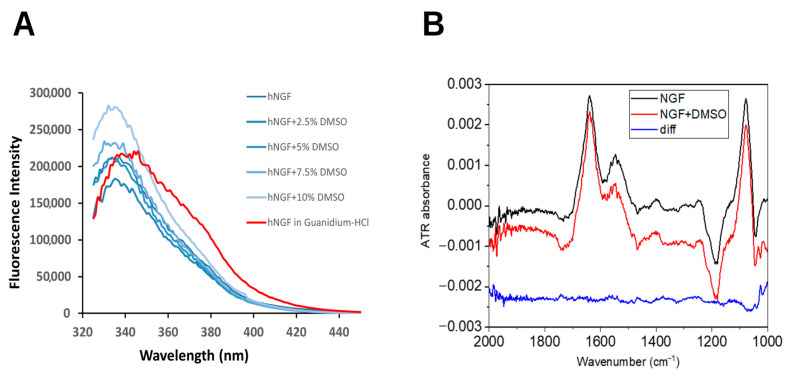
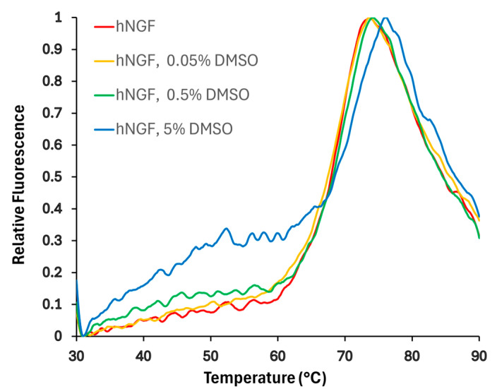
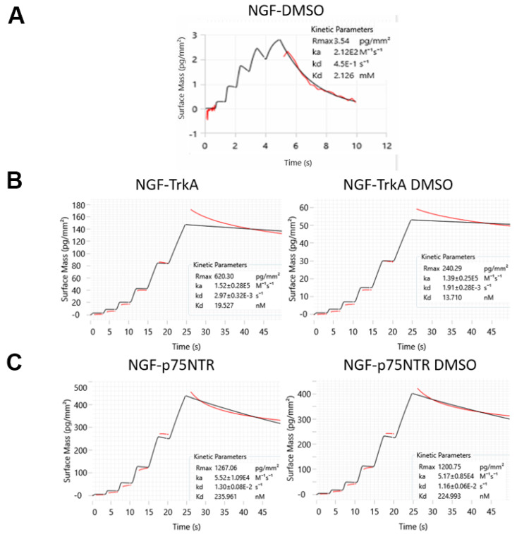
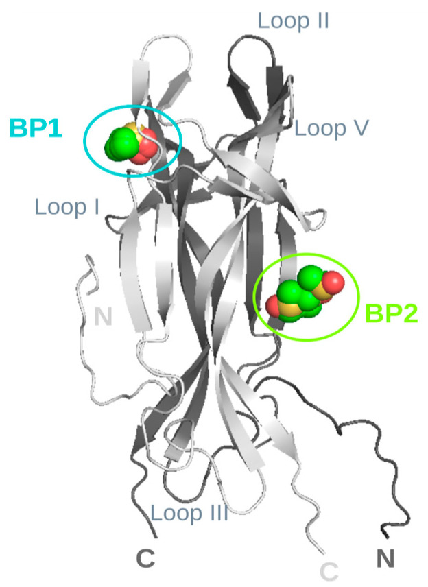
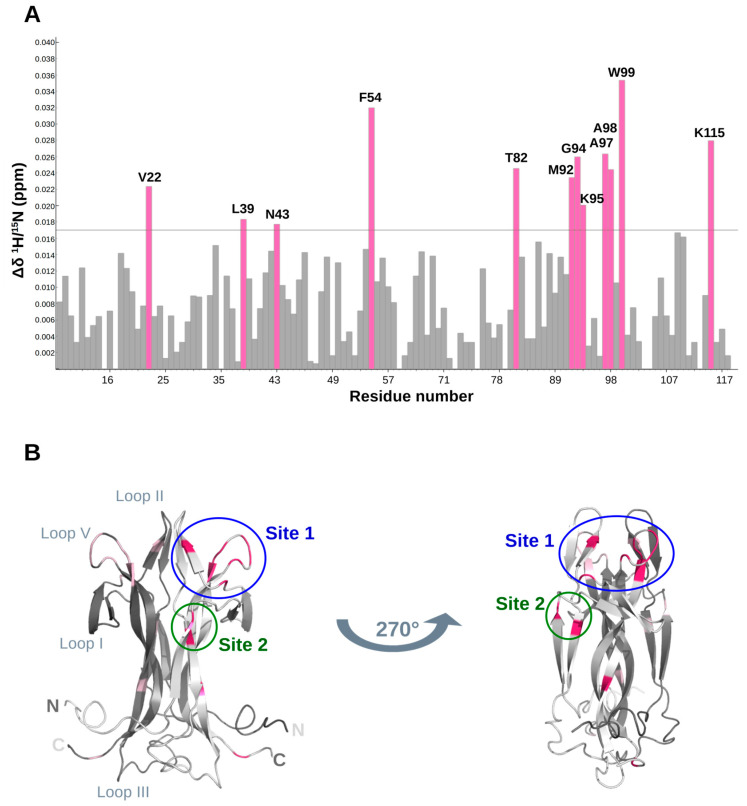
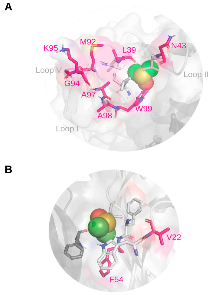
Background: The Nerve Growth Factor (NGF) is essential for neuronal survival and function and represents a key therapeutic target for pain and inflammation-related disorders, as well as for neurodegenerative diseases. Small-molecule antagonists of human NGF (hNGF) offer advantages over monoclonal antibodies, including oral availability and reduced immunogenicity. However, their development is often hindered by solubility challenges, necessitating the use of solvents like dimethyl sulfoxide (DMSO). This study investigates whether DMSO directly interacts with hNGF and affects its receptor-binding properties. Methods: Integrative/hybrid computational and experimental biophysical approaches were used to assess DMSO-NGF interaction by combining machine-learning tools and Nuclear Magnetic Resonance (NMR), Fourier Transform Infrared (FT-IR) spectroscopy, Differential Scanning Fluorimetry (DSF) and Grating-Coupled Interferometry (GCI). These techniques evaluated binding affinity, conformational stability, and receptor-binding dynamics. Results: Our findings demonstrate that DMSO binds hNGF with low affinity in a specific yet non-disruptive manner. Importantly, DMSO does not induce significant conformational changes in hNGF nor affect its interactions with its receptors. Conclusions: These results highlight the importance of considering solvent-protein interactions in drug discovery, as these low-affinity yet specific interactions can affect experimental outcomes and potentially alter the small molecules binding to the target proteins. By characterizing DMSO-NGF interactions, this study provides valuable insights for the development of NGF-targeting small molecules, supporting their potential as effective alternatives to monoclonal antibodies for treating pain, inflammation, and neurodegenerative diseases.

Keywords: Differential Scanning Fluorimetry (DSF); Fourier Transform Infrared (FT-IR) spectroscopy; Grating-Coupled Interferometry (GCI); HADDOCK; Nerve Growth Factor (NGF); Nuclear Magnetic Resonance (NMR); Protenix; dimethyl sulfoxide (DMSO); drug discovery; solvent effect.


Conflict of interest statement

The authors declare no conflicts of interest.

Figures

Figure 1
Figure 1
Impact of DMSO on hNGF structure. (A) Intrinsic fluorescence emission spectra of hNGF in the presence of increasing DMSO concentrations (from 2.5% to 10%, from dark to light blue). hNGF without DMSO: dark blue line. hNGF in 6 M Guanidine hydrochloride: red curve. (B) FT-IR spectra of hNGF in the presence and absence of DMSO. Blue spectrum: the difference between the red and black spectrum (downshifted for clarity). DMSO spectrum in Supplementary Figure S1.
Figure 2
Figure 2
Impact of DMSO concentration on hNGF melting curves obtained by DSF. Normalized curves are shown. The buffer conditions were 50 mM sodium phosphate, pH 6.6, and 50 mM NaCl. The effect of three different DMSO concentrations was tested, obtaining: Tm: 67.9 ± 0.1 °C (0% DMSO); 67.9 ± 0.5 °C (0.05% DMSO); 67.9 ± 0.4 °C (0.5% DMSO); 69.1 ± 0.9 °C (0.5% DMSO).
Figure 3
Figure 3
WaveRAPID kinetic and affinity data obtained using the Creoptix WAVE-delta system: (A) for hNGF-DMSO interaction and to compare hNGF binding to its receptors; (B) TrkA and (C) p75NTR, in the absence and presence of DMSO. All the quality assessments were fulfilled: χ2 (1.29 ± 0.15; 1.57 ± 0.34, 1.08 ± 0.22. 1.66 ± 0.28, and 1.13 ± 0.19 for hNGF-DMSO, hNGF-TrkA, hNGF-TrkA DMSO, hNGF- p75NTR, and hNGF-p75NTR DMSO, respectively); parameter errors and residual plots were acceptable, the sensorgrams had sufficient curvatures, and the kinetic constant kd was within a measurable range.
Figure 4
Figure 4
Ab initio models of hNGF-DMSO interaction. The identified binding pockets are labelled as BP1 (in cyan) and BP2 (in green), respectively, with loop regions, as well as N- and C-termini also annotated for reference. The two protomers of hNGF are coloured in light and dark grey, respectively. DMSO molecules representative of the two clusters are shown as spheres and colour-coded by element. Figure generated using PyMOL 2.1.0 [30].
Figure 5
Figure 5
Structural determinants of the hNGF-DMSO interaction. (A) DMSO-hNGF interactions by NMR titration. Map of combined 1H/15N Chemical Shift Perturbations (CSPs, Δδ) of the HN groups of hNGF upon DMSO binding. Residues exhibiting the largest CSP (Δδ > 0.017 ppm: threshold grey line shown in the Figure) are labelled in pink. (B) Mapping of CSP data at the endpoint of the DMSO/hNGF titration onto the 3D NMR solution structure of hNGF (PDB ID: 6YW8). Residues with significant CSP (Δδ > 0.017 ppm) are highlighted in pink. The two protomers of hNGF are coloured in light and dark grey, respectively. Two representations of hNGF are shown, with a 270° rotation along the z-axis, one with respect to the other. Proposed binding sites are indicated as Site 1 (in blue) and Site 2 (in green). Loop regions, as well as N- and C-termini also annotated for reference. Figure generated using PyMOL 2.1.0 [30].
Figure 6
Figure 6
Close-up views of the most significant representative HADDOCK docking poses of DMSO bound to hNGF. (A) Top view of the proposed Site 1 identified by CSP analysis, encompassing Loops II and V. (B) Side view of the proposed Site 2 identified by CSP analysis, involving the central stem region. The two protomers of hNGF are coloured in light and dark grey, respectively. The protein is displayed in cartoon representation, with an overlaid surface. Residues exhibiting the largest CSPs are shown as pink sticks and colour-coded by element. Hydrophobic residues of the binding site are depicted in dark grey and colour-coded by element. DMSO molecules are represented as spheres and colour-coded by element. Figure generated using PyMOL 2.1.0 [30].

References

    1. Levi-Montalcini R. The nerve growth factor 35 years later. Science. 1987;237:1154–1162. doi: 10.1126/science.3306916. - DOI - PubMed
    1. Levi-Montalcini R., Hamburger V. Selective growth stimulating effects of mouse sarcoma on the sensory and sympathetic nervous system of the chick embryo. J. Exp. Zool. 1951;116:321–361. doi: 10.1002/jez.1401160206. - DOI - PubMed
    1. Huang E.J., Reichardt L.F. Trk receptors: Roles in neuronal signal transduction. Annu. Rev. Biochem. 2003;72:609–642. doi: 10.1146/annurev.biochem.72.121801.161629. - DOI - PubMed
    1. Dechant G., Barde Y.-A. The neurotrophin receptor p75(NTR): Novel functions and implications for diseases of the nervous system. Nat. Neurosci. 2002;5:1131–1136. doi: 10.1038/nn1102-1131. - DOI - PubMed
    1. Chao M.V. Neurotrophins and their receptors: A convergence point for many signalling pathways. Nat. Rev. Neurosci. 2003;4:299–309. doi: 10.1038/nrn1078. - DOI - PubMed

MeSH terms

LinkOut - more resources