. 2025 Jun 30;13(7):714.
doi: 10.3390/vaccines13070714.

Replication of Vectored Herpesvirus of Turkey (HVT) in a Continuous, Microcarrier-Independent Suspension Cell Line from Muscovy Duck

Affiliations

Replication of Vectored Herpesvirus of Turkey (HVT) in a Continuous, Microcarrier-Independent Suspension Cell Line from Muscovy Duck

Karoline Mähl et al. Vaccines (Basel). .

Abstract

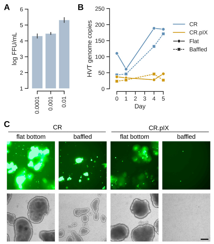
Background/Objectives: More than 33 billion chickens are industrially raised for meat and egg production globally and vaccinated against Marek's disease virus (MDV). The antigenically related herpesvirus of turkey (HVT) is used as a live-attenuated vaccine, commonly provided as a recombinant vector to protect chickens against additional unrelated pathogens. Because HVT replicates in a strictly cell-associated fashion to low levels of infectious units, adherent primary chicken or duck embryo fibroblasts are infected, dislodged from the cultivation surface and distributed as cryocultures in liquid nitrogen to the site of application. Although viable cells are complex products, application of infected cells in ovo confers protection even in presence of maternal antibodies. Methods/Results: The aim of our study was to determine whether a continuous cell line in a scalable cultivation format can be used for production of HVT-based vaccines. The AGE1.CR cell line (from Muscovy duck) was found to be highly permissive in adherent cultures. Propagation in suspension, however, initially gave very low yields. The induction of cell-to-cell contacts in carrier-independent suspensions and a metabolic shock improved titers to levels suitable for vaccine production (>105 infectious units/mL after infection with multiplicity of 0.001). Conclusions: Production of HVT is challenging to scale to large volumes and the reliance on embryonated eggs from biosecure facilities is complex. We demonstrate that a cell-associated HVT vector can be propagated in a carrier-independent suspension culture of AGE1.CR cells in chemically defined medium. The fed-batch production is independent of primary cells and animal-derived material and can be scaled to large volumes.

Keywords: HVT; MDV; Marek’s disease vaccine; suspension cell culture.


Conflict of interest statement

Authors K.M., S.A., D.H., A.K., V.S. and I.J. are employed by a company (ProBioGen AG) involved in vaccine research and development.

Figures

Figure 1
Figure 1
Uninfected cells were either pre-seeded one day prior to infection or co-seeded together with infected cells. The fluorescent foci were quantified by automated analysis with a plate reader. (A) Focus diameter (in µm) expand with a similar kinetic in both CR and CS cells independent of infection format. The usage of CR cells generally leads to larger (fluorescent) foci areas (in mm2) (B) and shows a higher number of foci (C) than CS cells. Although CR cell focus formation also improves by co-seeding, the effect is stronger for CS cells. Each curve is the mean of two independent experiments with seeding at higher cell density. (D) The effects appear not to be a chance event and were observed with different preparations of seed virus (23H08O and 24H12F; (AC) were performed with the latter preparation) for both cell lines. (E) Example of foci obtained in CR and CS cell lines with either pre- or co-seeding. Foci generally appear to be more compact for CR cells than CS cells. Infection was performed with 100 FFU per well of a 12-well plate. Without uninfected cells, the seed did not expand into foci. The scale bar corresponds to 250 µm.
Figure 2
Figure 2
Endpoint observations at day 7 after infection by pre- or co-seeding with 100 FFU per well of a 12-well plate. (A) Co-seeding leads to higher number of foci also for the CR- and CS-derived cell lines that express the pIX protein. DF-1 cells are barely susceptible to HVT-GFP and only 2 foci were obtained by co-seeding. (B) Focus diameters (in µm) show similar expansion at day 7 but are surprisingly low for CR cells. This effect may possibly be caused by loss of some expanded foci to cytopathic effect because of the long incubation until day 7 (see also Figure 3). The error bars show standard deviation based on number of foci detected per respective well shown in (A). (C) Genome copy numbers for HVT are consistent with the observed number of foci. Because the DF-1 cell line does not encode the E1A gene that was used to immortalize the CR cell line [21], absolute numbers are given here for HVT and (where appropriate, hatched columns) for E1A. (D) Genome copy numbers normalized to the cellular E1A reference. Absolute copy numbers are shown on a log scale, all other values on a linear scale. Results shown here are the averages of two independent experiments.
Figure 3
Figure 3
Number of foci (A) and size of foci in µm (B) compared between day 3 and day 7 after infection. The bold line in the violin plot depicts the median value, the thin lines the upper and lower quartiles. Appearance and development of foci from day 3 to day 7 (C). The arrow at day 4 in the row with CR cells points to a focus that developed into a true plaque where cells in the center dislodge. Such plaques are usually regrown with fresh cells that are also infected. The scale bar corresponds to 250 µm.
Figure 4
Figure 4
(A) Suspension CR cells in a non-baffled 125 mL-shaker flask were infected with MOIs from of 0.0001 to 0.01 and infectious titers were determined for samples at day 7 post-infection. Optimization of the suspension process was continued with the central MOI of 0.001. In (B) and (C), CR and CR.pIX infection was compared in baffled and flat-bottom shaker flasks. (B) Genome copy numbers (sORF1 copies normalized to E1A copies) increased during the infection in CR cells but remained essentially constant at the input level for CR.pIX. (C) While EGFP signals were strong in aggregates in CR cells infected in flat-bottom shaker flasks, aggregates were fewer and smaller in baffled flasks and EGFP signals less pronounced. CR.pIX cells did not perform well even in non-baffled shaker flasks. The scale bar corresponds to 250 µm.
Figure 5
Figure 5
Sudden depletion of IGF improves titers significantly. (A) EGFP signal increased especially strongly in aggregates of CR and CR.pIX cells in the shock-depleted cultures. The first panel shows cultures 1 day after infection, the center panel at day 5 and the bottom panel the harvest also at day 5 after dissociation of aggregates. The scale bar corresponds to 250 µm. (B) Viral genome copy numbers (sORF1 copies normalized to E1A) increased to highest levels in shock-depleted CR cultures, followed by normal CR and shock-depleted CR.pIX cultures. (C) Viral genome copy numbers were determined by ddPCR against the EGFP transgene and the viral sORF1 gene. Ratios of sORF1 to EGFP were approximately 3 throughout the cultures in both cell lines. This ratio is consistent with non-fluorescent to fluorescent foci in titrations. (D) Infectious titers in FFU/mL obtained with the dissociated aggregates. The infectious titers may be underestimated by a factor of 3.
Figure 6
Figure 6
The suspension process tolerates differences in production media composition and ratios. (A) EGFP signal distribution and aggregate appearances at the day of harvest prior to dissociation. Ratios (such as 1:1) refer to amount production medium added relative to the suspension medium CD-U7. The bottom panel depicts the experiment where no production medium (only 1 volume CD-U7) was added. The scale bar corresponds to 250 µm. (B) Genome copy numbers increase, especially starting at day 3. Values were best for DMEM as production medium or if DMEM/F12 is given at least at 1:1. (C) Infectious titers at day 5 were above 105 FFU/mL for the three cultures that exhibited strong genome copy number increases. Aggregate induction is important as shown by infectious titers in the CD-U7 only culture that correspond to less than input virus (infection with MOI 0.001 × 2 × 106 cells/mL at the time of infection equals 2 × 10³ FFU/mL).

Similar articles

References

    1. Sheppard M. Viral Vectors for Veterinary Vaccines. Adv. Vet. Med. 1999;41:145. - PMC - PubMed
    1. Okura T., Taneno A., Oishi E. Cell-to-Cell Transmission of Turkey Herpesvirus in Chicken Embryo Cells via Tunneling Nanotubes. Avian Dis. 2021;65:335–339. doi: 10.1637/aviandiseases-D-21-00022. - DOI - PubMed
    1. Okura T., Otomo H., Taneno A., Oishi E. Replication Kinetics of Turkey Herpesvirus in Lymphoid Organs and Feather Follicle Epithelium in Chickens. J. Vet. Med. Sci. 2021;83:1582–1589. doi: 10.1292/jvms.21-0247. - DOI - PMC - PubMed
    1. Palya V., Tatár-Kis T., Mató T., Felföldi B., Kovács E., Gardin Y. Onset and Long-Term Duration of Immunity Provided by a Single Vaccination with a Turkey Herpesvirus Vector ND Vaccine in Commercial Layers. Vet. Immunol. Immunopathol. 2014;158:105–115. doi: 10.1016/j.vetimm.2013.11.008. - DOI - PubMed
    1. Fiddaman S.R., Dimopoulos E.A., Lebrasseur O., du Plessis L., Vrancken B., Charlton S., Haruda A.F., Tabbada K., Flammer P.G., Dascalu S., et al. Ancient Chicken Remains Reveal the Origins of Virulence in Marek’s Disease Virus. Science. 2023;382:1276–1281. doi: 10.1126/science.adg2238. - DOI - PubMed

LinkOut - more resources