Review
. 2025 Jul 20;13(7):767.
doi: 10.3390/vaccines13070767.

Bioengineering Outer-Membrane Vesicles for Vaccine Development: Strategies, Advances, and Perspectives

Affiliations
Review

Bioengineering Outer-Membrane Vesicles for Vaccine Development: Strategies, Advances, and Perspectives

Ayesha Zahid et al. Vaccines (Basel). .

Abstract

Outer-membrane vesicles (OMVs), naturally secreted by Gram-negative bacteria, have gained recognition as a versatile platform for the development of next-generation vaccines. OMVs are essential contributors to bacterial pathogenesis, horizontal gene transfer, cellular communication, the maintenance of bacterial fitness, and quorum sensing. Their intrinsic immunogenicity, adjuvant properties, and scalability establish OMVs as potent tools for combating infectious diseases and cancer. Recent advancements in genetic engineering and biotechnology have further expanded the utility of OMVs, enabling the incorporation of multiple epitopes and antigens from diverse pathogens. These developments address critical challenges such as antigenic variability and co-infections, offering broader immune coverage and cost-effective solutions. This review explores the unique structural and immunological properties of OMVs, emphasizing their capacity to elicit robust immune responses. It critically examines established and emerging engineering strategies, including the genetic engineering of surface-displayed antigens, surface conjugation, glycoengineering, nanoparticle-based OMV engineering, hybrid OMVs, and in situ OMV production, among others. Furthermore, recent advancements in preclinical research on OMV-based vaccines, including synthetic OMVs, OMV-based nanorobots, and nanodiscs, as well as emerging isolation and purification methods, are discussed. Lastly, future directions are proposed, highlighting the potential integration of synthetic biology techniques to accelerate research on OMV engineering.

Keywords: bioengineered vaccines; cancer vaccines; genetic engineering; multi-antigen vaccines; multi-pathogen vaccines; outer membrane vesicles; vaccine development.


Conflict of interest statement

The authors declare that this research was conducted in the absence of any commercial or financial relationships that could be construed as potential conflicts of interest.

Figures

Figure 1
Figure 1
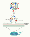
OMVs: Small packages with large roles in bacterial physiology. OMVs originate from the outer membrane of parent bacteria and inherently encapsulate numerous cellular substances. The bio-functions of OMVs are classified and delineated as follows: toxin secretion, horizontal DNA transfer, antimicrobial delivery, immunomodulation, nutrition digestion, quorum sensing, and secretion of misfolded proteins.
Figure 2
Figure 2
Biogenesis and composition of OMVs. An OMV comprises an inner membrane and an outer membrane. Phospholipids, lipopolysaccharides, and outer-membrane proteins are present in the outer membrane. Phospholipids and integral membrane proteins are prevalent in the inner membrane. OMVs carry internal constituents such as DNA and RNA, along with outer-membrane proteins and components from the periplasmic space.
Figure 3
Figure 3
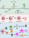
Harnessing OMVs for vaccination: bridging innate and adaptive immunity. (A) Identification of OMV-related pathogen-associated molecular patterns (PAMPs) by pattern recognition receptors (PRRs). Arrows denote ligand–receptor interactions between PAMPs and NOD-like receptors (NLRs) or Toll-like receptors (TLRs). (B) Upon delivery, OMV vaccines, including antigens and diverse PAMPs, are identified and engulfed by immature dendritic cells after the interaction of PAMPs with PRRs. The identification and encapsulation of OMV vaccines by dendritic cells promote their development, characterized by the production of co-stimulatory molecules and cytokine release, as well as antigen presentation. (C) Mature dendritic cells presenting bacterial antigens stimulate the activation and proliferation of antigen-specific CD4+ and CD8+ T lymphocytes in lymph nodes. In response to distinct cytokine environments, CD4+ T-cells differentiate into Th1 and Th2 cells. Th2 cells assist B-cells in generating antigen-specific antibodies that adhere to, and facilitate the elimination of, germs through opsonization. CD8+ cytotoxic T lymphocytes (CTLs) identify and eliminate bacteria-infected cells by exerting cytotoxic effects and releasing cytokines, including IFN-γ, upon encountering bacterial antigens. The efficacy of CTLs is augmented by Th1 cells via the secretion of IFN-γ. B-cell: B lymphocyte (Bursa-derived cell); CpG: Cytosine-phosphate-Guanine (CpG) dinucleotides; CD4+ T-cells: Cluster of differentiation four positive T-cells; CD8+ T-cells: Cluster of differentiation eight positive T-cells; IFN-γ: Interferon gamma; NOD-1/2: NOD-like receptor 1/2; TLR-2: Toll-like receptor 2; TLR-4: Toll-like receptor 4; TLR-5: Toll-like receptor 5; TLR-9: Toll-like receptor 9; T-cell: T lymphocyte (Thymus-derived cell); Th1 cell: T helper 1 cell; Th2 cell: T helper 2 cell; OMV: outer-membrane vesicle.
Figure 4
Figure 4
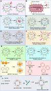
OMVs are a versatile tool for genetic modification and bioengineering in the design of effective vaccines. OMVs can be bioengineered for the following purposes: (A) Minimizing the ability of LPS to trigger a reactogenic response following OMV injection [189,190,191]. (B) Enhancing the natural ability of Gram-negative bacteria to release OMVs [192]. (C) Expressing antigens in the lumen of OMVs [193]. (D) Expressing various protein or peptide antigens on the surface through genetically modifying the parent bacteria [186]. (E) Linking externally purified protein antigens with the surface proteins of OMVs [194,195]. (F) Linking externally purified protein antigens with the surface LPS of OMVs [196]. (G) Conjugating polysaccharide-based antigens through fusion with surface proteins or LPS [197,198]. (H) Genetically modifying the parent bacteria to express polysaccharide antigens [199]. (I) Coating nanoparticles (NPs) with OMVs [200]. (J) Preparing hybrid OMVs through the fusion of OMVs with tumour or plant membranes or liposomes [201]. (K) SpyTag/SpyCatcher system for antigen display [202]. LPS: lipopolysaccharide; NP: nanoparticles; OMV: outer-membrane vesicle.
Figure 5
Figure 5
In situ release of engineered outer-membrane vesicles (OMVs) from transplanted bacteria. Genetically modified bacteria can be administered to a host to actively generate and release OMVs in situ. These OMVs subsequently deliver their surface-associated and internal biomolecules into the membranes or cytoplasm of target cells, facilitating precise biological functions.
Figure 6
Figure 6
Preparation and engineering of OM-NDs and OMV-based nanorobots. (A) Outer-membrane vesicles (OMVs) derived from Pseudomonas aeruginosa are isolated and treated with styrene-maleic acid (SMA), resulting in the formation of OM-NDs. This formulation serves as a potential nano-vaccine for protection against bacterial infection. The figure is adapted from [355]. (B) Fabrication and characterization of OMV-siRNA robots. The panel depicts the fabrication process of OMV-siRNA nanorobots, featuring surface-engineered CPP designed for targeted tumour binding and penetration. Biocatalytic propulsion improves the targeted binding and penetration of siRNA-loaded OMV nanorobots (OMV-siR robots) at the tumour site. The figure is adapted from [356]. ClyA-CPP: Cytolysin A-fused cell-penetrating peptide; siRNA: Small interfering RNA; OM-NDs: Outer-membrane vesicle-based nanodisc; OMV: outer-membrane vesicle.
Figure 7
Figure 7
Synthetic biology strategies for engineering outer-membrane vesicles (OMVs). (A) Potential synthetic biology-based engineering strategies that can be applied to modify OMV-producing strains. These modifications aim to enhance OMV production and optimize the therapeutic cargo of microbially derived OMVs. (B) Schematic representation of the cell-free synthesis of OMV components, utilizing cell-free protein synthesis for OMV assembly. (C) Overview of strain-engineering approaches for OMV production, including CRISPR/Cas9-based gene regulation, knockout and knock-in strategies, and inducible gene expression to modulate OMV yield and content. (D) For OMV cargo engineering, xeno nucleic acids, unnatural amino acids, and metabolic engineering can be incorporated to generate antigens for therapeutic and vaccine applications. AI, artificial intelligence; CDS: coding sequence; CRISPR/dCas9: clustered regularly interspaced short palindromic repeats (CRISPR)/endonuclease deficient CRISPR-associated protein 9 (dCas9); OMV: outer-membrane vesicle.

Similar articles

References

    1. Schwechheimer C., Kuehn M.J. Outer-membrane vesicles from Gram-negative bacteria: Biogenesis and functions. Nat. Rev. Microbiol. 2015;13:605–619. doi: 10.1038/nrmicro3525. - DOI - PMC - PubMed
    1. Turnbull L., Toyofuku M., Hynen A.L., Kurosawa M., Pessi G., Petty N.K., Osvath S.R., Cárcamo-Oyarce G., Gloag E.S., Shimoni R. Explosive cell lysis as a mechanism for the biogenesis of bacterial membrane vesicles and biofilms. Nat. Commun. 2016;7:11220. doi: 10.1038/ncomms11220. - DOI - PMC - PubMed
    1. Bishop D., Work E. An extracellular glycolipid produced by Escherichia coli grown under lysine-limiting conditions. Biochem. J. 1965;96:567. doi: 10.1042/bj0960567. - DOI - PMC - PubMed
    1. Knox K., Vesk M., Work E. Relation between excreted lipopolysaccharide complexes and surface structures of a lysine-limited culture of Escherichia coli. J. Bacteriol. 1966;92:1206–1217. doi: 10.1128/jb.92.4.1206-1217.1966. - DOI - PMC - PubMed
    1. Rothfield L., Pearlman-Kothencz M. Synthesis and assembly of bacterial membrane components: A lipopolysaccharide-phospholipid-protein complex excreted by living bacteria. J. Mol. Biol. 1969;44:477–492. doi: 10.1016/0022-2836(69)90374-X. - DOI - PubMed

LinkOut - more resources