. 2025 Jul 30:19476035251361679.
doi: 10.1177/19476035251361679. Online ahead of print.

miR-708-5p Attenuates Osteoarthritis Progression via Multi-Target Modulation of the NOX4/NF-κB Axis and Cartilage Homeostasis

Affiliations

miR-708-5p Attenuates Osteoarthritis Progression via Multi-Target Modulation of the NOX4/NF-κB Axis and Cartilage Homeostasis

Shih-Hao Huang et al. Cartilage. .

Abstract

ObjectiveTo investigate the novel role of miR-708-5p in osteoarthritis (OA) and its potential as a therapeutic target through regulation of NOX4/NF-κB signaling.MethodsExpression levels of miR-708-5p were analyzed in OA cartilage using GEO datasets and validated in interleukin (IL)-1β-treated primary human chondrocytes. Gain- and loss-of-function experiments were performed using miR-708-5p mimics and inhibitors to evaluate its effects on inflammation, extracellular matrix metabolism, apoptosis, and oxidative stress. Direct targeting of NOX4 by miR-708-5p was confirmed through bioinformatic prediction, luciferase reporter assays, and rescue experiments.ResultsmiR-708-5p was significantly downregulated in OA cartilage and IL-1β-treated chondrocytes. Overexpression of miR-708-5p attenuated IL-1β-induced inflammatory responses by suppressing pro-inflammatory cytokines (IL-1β, IL-6, tumor necrosis factor [TNF]-α), inhibiting matrix-degrading enzymes (MMP3, ADAMTS-4), and enhancing anabolic factors (COL2A1, SOX9). miR-708-5p protected against chondrocyte apoptosis by regulating Bcl2/BAX and caspase-3 expression. It also increased chondrocyte proliferation in EdU assays and reduced reactive oxygen species (ROS) production. Mechanistically, miR-708-5p directly inhibited NOX4, reducing ROS generation and nuclear factor kappa B (NF-κB) activation. NOX4 overexpression reversed the protective effects of miR-708-5p, confirming the functional significance of this regulatory axis.ConclusionmiR-708-5p is downregulated in OA and exerts chondroprotective effects. These findings suggest that restoring miR-708-5p expression may effectively suppress the NOX4/NF-κB axis and modulate chondrocyte inflammation, oxidative stress, apoptosis, and matrix degradation.

Keywords: NOX4/NF-κB signaling; cartilage homeostasis; chondrocyte inflammation; miR-708-5p; osteoarthritis.


Conflict of interest statement

The author(s) declared no potential conflicts of interest with respect to the research, authorship, and/or publication of this article.

Figures

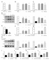
“Statistical analysis reveals significant upregulation of miR-708-5p in OA tissues compared to controls. GSE105027, GSE143514. Deteriorated miR-708-5p expression over time correlates with decreased ODz4 expression in IL-1β-treated cells.”
Figure 1.
miR-708-5p expression is downregulated in osteoarthritic cartilage and IL-1β-stimulated chondrocytes. (A, B) Expression levels of miR-708-5p in normal and osteoarthritis (OA) cartilage tissues from GEO datasets (A) GSE105027 and (B) GSE143514. (C, D) Time-dependent suppression of (C) miR-708-5p and its host gene (D) Odz4 expression in human chondrocytes following stimulation with IL-1β (10 ng/ml) for indicated periods (0, 1, 6, 12, or 24 hours). Expression levels were determined by real-time PCR and normalized to GAPDH. Data are presented as mean ± SD. Statistical significance: * P < 0.05, ** P < 0.01, *** P < 0.001 compared to control group.
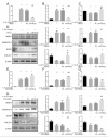
The image depicts a study analyzing the effects of miR-708-5p on IL-1β-induced inflammatory responses in human chondrocytes. The study includes RT-qPCR analysis of miR-708-5p expression, effects on inflammatory mediators, and ELISA analysis.
Figure 2.
miR-708-5p inhibits IL-1β-induced inflammatory responses in human chondrocytes. (A) RT-qPCR analysis of miR-708-5p expression in chondrocytes transfected with miR-708-5p mimics or negative control (NC). (B-D) Effects of miR-708-5p overexpression on inflammatory mediators. RT-qPCR analysis of (B) IL-1β and (C) IL-6 mRNA expressions. (D) Western blot analysis and quantification of IL-6 and TNF-α protein levels in chondrocytes transfected with miR-708-5p mimics or NC and treated with IL-1β (10 ng/ml) for 24h. β-Actin served as loading control. (E) RT-qPCR analysis of miR-708-5p expression in chondrocytes transfected with miR-708-5p inhibitor or inhibitor negative control (NC). (F-H) Effects of miR-708-5p inhibition on inflammatory mediators. RT-qPCR analysis of (F) IL-1β and (G) IL-6 mRNA expressions. (H) Western blot analysis and quantification of IL-6 and TNF-α protein levels in chondrocytes transfected with miR-708-5p inhibitor or NC and stimulated with IL-1β (10 ng/ml) for 24h. (I-L) ELISA analysis of IL-6 and TNF-α production in culture supernatants. Concentration of (I, K) IL-6 and (J, L) TNF-α in supernatants from chondrocytes transfected with (I, J) miR-708-5p mimics or (K, L) miR-708-5p inhibitor and stimulated with IL-1β (10 ng/ml) for 24h. Data are presented as mean ± SD. Statistical significance: * P < 0.05, ** P < 0.01, *** P < 0.001 compared to untreated control; # P < 0.05, ## P < 0.01, ### P < 0.001 compared to IL-1β + NC group.
The image depicts a scientific study on the regulation of extracellular matrix homeostasis in chondrocytes by miR-708-5p through RT-qPCR analysis, Western blot analysis, and quantitative protein measurements. The study involves comparing the effects of miR-708-5p mimics, miR-708-5p inhibitor, and various treatments on the expression of MMP3, ADAMTS-4, SOX9, and COL2A1 in chondrocytes. The results are shown in a series of bar graphs and protein visualization images with statistical significance markers, indicating the impact of IL-1β and miR-708-5p manipulation on these genes and proteins.
Figure 3.
miR-708-5p regulates extracellular matrix homeostasis in IL-1β-stimulated chondrocytes. (A-C) RT-qPCR analysis of mRNA expression for (A) MMP3, (B) ADAMTS4, and (C) COL2A1 in chondrocytes transfected with miR-708-5p mimics and stimulated with IL-1β (10 ng/ml) for 24h. (D) Western blot analysis and densitometric quantification of catabolic (MMP3, ADAMTS-4) and anabolic (SOX9, COL2A1) proteins in chondrocytes transfected with miR-708-5p mimics or negative control (NC) and stimulated with IL-1β (10 ng/ml) for 24h. β-Actin served as loading control. (E-G) RT-qPCR analysis of mRNA expression for (E) MMP3, (F) ADAMTS-4, and (G) COL2A1 in chondrocytes transfected with miR-708-5p inhibitor and stimulated with IL-1β (10 ng/ml) for 24h. (H) Western blot analysis and quantification of MMP3, ADAMTS-4, SOX9, and COL2A1 proteins in chondrocytes transfected with miR-708-5p inhibitor or inhibitor NC and stimulated with IL-1β (10 ng/ml) for 24h. Data are presented as mean ± SD. * P < 0.05, ** P < 0.01, *** P < 0.001 compared to untreated control; # P < 0.05, ## P < 0.01, ### P < 0.001 compared to IL-1β + NC group.
The image shows a series of graphs and Western blot images evaluating the effects of miR-708-5p on IL-1β-induced chondrocyte apoptosis. The graphs display RT-qPCR and Western blot data for Bcl2 and caspase-3 expression levels. The image also includes a Western blot showing the levels of BAX, Cleaved Caspase3, and β-Actin in chondrocytes transfected with miR-708-5p, miR708 inhibitor, and negative control. The data suggests different levels of mRNA and protein expression under various conditions.
Figure 4.
miR-708-5p modulates IL-1β-induced chondrocyte apoptosis. (A-B) RT-qPCR analysis of mRNA expression for (A) Bcl2 and (B) caspase-3 in chondrocytes transfected with miR-708-5p mimics or negative control (NC) and treated with IL-1β (10 ng/ml) for 24h. (C) Western blot analysis and densitometric quantification of apoptotic markers Bcl-2-associated X protein (BAX) and cleaved caspase-3 in chondrocytes transfected with miR-708-5p mimics or NC and stimulated with IL-1β (10 ng/ml) for 24h. β-Actin served as loading control. (D-E) RT-qPCR analysis of mRNA expression for (D) Bcl2 and (E) caspase-3 in chondrocytes transfected with miR-708-5p inhibitor or NC and treated with IL-1β (10 ng/ml) for 24h. (F) Western blot analysis and quantification of BAX and cleaved caspase-3 proteins in chondrocytes transfected with miR-708-5p inhibitor or NC and stimulated with IL-1β (10 ng/ml) for 24h. Data are presented as mean ± SD. * P < 0.05, ** P < 0.01, *** P < 0.001 compared to untreated control; # P < 0.05, ## P < 0.01, ### P < 0.001 compared to IL-1β + NC group.
In the provided images, human chondrocytes were transfected with either miR-708-5p mimics or inhibitor, and subsequently treated with IL-1B for 24 hours to assess cell proliferation using EdU incorporation assay. The experiment showed a significant inhibition of cell proliferation by miR-708-5p compared to all controls, as demonstrated by the lower percentage of EdU-positive cells in the graphs. This suggests that miR-708-5p negatively regulates chondrocyte proliferation under IL-1B stimulation.
Figure 5.
miR-708-5p regulates chondrocyte proliferation under IL-1β stimulation. (A-B) Human chondrocytes were transfected with (A) miR-708-5p mimics or (B) miR-708-5p inhibitor or respective negative controls (NC) for 24h, then treated with IL-1β (10 ng/ml) for 24h. Cell proliferation was assessed by EdU incorporation assay (green) with DAPI nuclear counterstaining (blue). Quantification is shown as the percentage of EdU-positive cells (right panels). ***P < 0.001 compared to untreated control; ## P < 0.01, ### P < 0.001 compared to IL-1β + respective NC group.
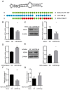
The image presents a detailed experimental analysis of miR-708-5p, a microRNA that targets NOX4, a gene involved in inflammatory processes. The sequence alignment shows miR-708-5p specifically binds to the 3'UTR of NOX4, altering its expression. NOX4 expression is significantly upregulated in cells co-expressed with miR-708-5p mimic, reduced with its inhibitor. Western blot analyses confirm these findings at the protein level. The luciferase reporter assays validate that miR-708-5p suppresses NOX4 by binding to its 3'UTR, as the activity is markedly lower with the mutant version.
Figure 6.
NOX4 is a direct target of miR-708-5p. (A) Predicted secondary structure of miR-708-5p and sequence alignment showing the binding site between miR-708-5p and NOX4 3'UTR. The wild-type (WT) sequence and mutated (MUT) binding site are shown with mutated nucleotides highlighted in red. (B-C) Effects of miR-708-5p on NOX4 expression: (B) Relative NOX4 mRNA expression and (C) representative Western blot with quantification of NOX4 protein expression in chondrocytes transfected with miR-708-5p mimic or negative control (NC). (D-E) Effects of miR-708-5p inhibition on NOX4 expression: (D) Relative NOX4 mRNA expression and (E) representative Western blot with quantification of NOX4 protein expression in chondrocytes transfected with miR-708-5p inhibitor or negative control (NC). β-Actin served as loading control for all Western blots. (F-G) Luciferase reporter assay results showing relative luciferase activity in chondrocytes co-transfected with miR-708-5p mimic or NC along with (F) wild-type NOX4 3'UTR or (G) mutated NOX4 3'UTR reporter constructs. **P < 0.01, ***P < 0.001 compared to respective NC.
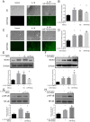
The image presents a scientific study examining the effects of miR-708-5p on IL-1β-induced oxidative stress and NOX4/NF-κB signaling.
Figure 7.
miR-708-5p modulates IL-1β-induced oxidative stress and NOX4/NF-κB signaling in chondrocytes. (A-D) Effects of miR-708-5p on ROS production and NOX4 expression. Human chondrocytes were transfected with miR-708-5p (A-B) mimics or (C-D) inhibitors and treated with IL-1β (10 ng/ml) for 24h. (A, C) ROS production visualized with DCFH-DA staining (green fluorescence, bottom panels) and corresponding phase contrast images (top panels). (B, D) RT-qPCR analysis of NOX4 mRNA expression under the same treatment conditions. (E-F) Western blot analysis and quantification of NOX4 protein expression in IL-1β-stimulated chondrocytes after transfection with miR-708-5p (E) mimics or (F) inhibitor, with β-Actin as loading control. (G-H) Western blot analysis and quantification of NF-κB pathway activation showing phosphorylated NF-κB (p-NF-κB) and total NF-κB in IL-1β-stimulated chondrocytes transfected with miR-708-5p (G) mimics or (H) inhibitor relative to appropriate controls. * P < 0.05, ** P < 0.01 compared to untreated control; # P < 0.05, ## P < 0.01 compared to IL-1β + respective negative control (NC) group.
The image shows an experiment investigating the effects of miR-708-5p and NOX4 on IL-1β-induced inflammatory and catabolic responses in human chondrocytes. Western blot analysis (A) reveals NOX4 protein expression increases in chondrocytes treated with NOX4 overexpression plasmid. Cell viability assay (B) shows a 10% decrease in cell viability with NOX4 overexpression. ELISA analysis (C-D) indicates 500% increase in IL-6 and 900% increase in TNF-α production with NOX4 overexpression. The study suggests that miR-708-5p and NOX4 play a significant role in modulating IL-1β-induced inflammatory responses in chondrocytes.
Figure 8.
The miR-708-5p/NOX4/NF-κB axis modulates IL-1β-induced inflammatory and catabolic responses in chondrocytes. (A) Western blot analysis and densitometric quantification of MMP3, MMP13, iNOS, NOX4, and caspase3 protein expression in human chondrocytes. Cells were co-transfected with miR-708-5p mimics and/or NOX4 overexpression plasmid (pcNOX4) as indicated, then stimulated with IL-1β (10 ng/ml) for 24h. β-Actin served as loading control. (B) Cell viability assessed by MTT assay in chondrocytes under the same treatment conditions as in (A). Results expressed as OD570 nm. (C-D) ELISA analysis of (C) IL-6 and (D) TNF-α production in culture supernatants of chondrocytes treated as in (A). ** P < 0.01, *** P < 0.001 compared to untreated control; # P < 0.05, ### P < 0.001 compared to IL-1β treated group; & P < 0.05, && P < 0.01, *** P < 0.001 compared to IL-1β + miR708-5p group.

Similar articles

References

    1. Glasson SS, Askew R, Sheppard B, Carito B, Blanchet T, Ma HL, et al. Deletion of active ADAMTS5 prevents cartilage degradation in a murine model of osteoarthritis. Nature. 2005;434(7033):644-8. doi: 10.1038/nature03369. - DOI - PubMed
    1. Aicher WK, Rolauffs B. The spatial organisation of joint surface chondrocytes: review of its potential roles in tissue functioning, disease and early, preclinical diagnosis of osteoarthritis. Ann Rheum Dis. 2014;73(4):645-53. doi: 10.1136/annrheumdis-2013-204308. - DOI - PubMed
    1. Hunter DJ, Bierma-Zeinstra S. Osteoarthritis. Lancet. 2019;393(10182):1745-59. doi: 10.1016/S0140-6736(19)30417-9. - DOI - PubMed
    1. Roemer FW, Kwoh CK, Hayashi D, Felson DT, Guermazi A. The role of radiography and MRI for eligibility assessment in DMOAD trials of knee OA. Nat Rev Rheumatol. 2018;14(6):372-80. doi: 10.1038/s41584-018-0010-z. - DOI - PMC - PubMed
    1. van Lieshout WAM, Valkering KP, Koenraadt KLM, van Etten-Jamaludin FS, Kerkhoffs GMMJ, van Geenen RCI. The negative effect of joint line elevation after total knee arthroplasty on outcome. Knee Surg Sports Traumatol Arthrosc. 2019;27(5):1477-86. doi: 10.1007/s00167-018-5099-8. - DOI - PMC - PubMed

LinkOut - more resources