Review
. 2025 Jul 28:8:25152564251363050.
doi: 10.1177/25152564251363050. eCollection 2025 Jan-Dec.

Membrane Contact Sites in Proteostasis and ER Stress Response

Affiliations
Review

Membrane Contact Sites in Proteostasis and ER Stress Response

Febe Vermue et al. Contact (Thousand Oaks). .

Abstract

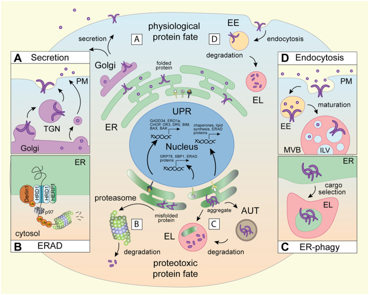
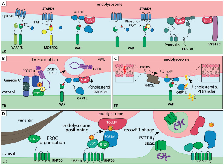
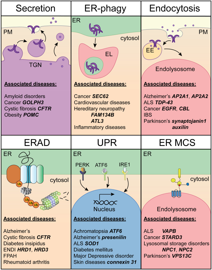
Execution of all cellular functions depends on a healthy proteome, whose maintenance requires multimodal oversight. Roughly a third of human proteins reside in membranes and thus present unique topological challenges with respect to biogenesis and degradation. To meet these challenges, eukaryotes have evolved organellar pathways of protein folding and quality control. Most transmembrane proteins originate in the endoplasmic reticulum (ER), where they are subject to surveillance and, if necessary, removal through either ER-associated proteasomal degradation (cytosolic pathway) or selective autophagy (ER-phagy; organellar pathway). In the latter case, ER cargoes are shuttled to (endo)lysosomes - the same organelles that degrade cell surface molecules via endocytosis. Here, we provide an overview of dynamic coordination between the ER and endolysosomes, with a focus on their engagement in specialized physical interfaces termed membrane contact sites (MCSs). We cover how cross-compartmental integration through MCSs allows biosynthetic and proteolytic organelles to fine-tune each other's membrane composition, organization, and dynamics and facilitates recovery from proteotoxic stress. Along the way, we highlight recent developments and open questions at the crossroads between organelle biology and protein quality control and cast them against the backdrop of factor-specific diseases associated with perturbed membrane homeostasis.

Keywords: endolysosome; endoplasmic reticulum; membrane contact sites; proteostasis; proteotoxic stress.


Conflict of interest statement

The authors declared no potential conflicts of interest with respect to the research, authorship, and/or publication of this article.

Figures

Figure 1.
Figure 1.
Biosynthetic and proteolytic membrane pathways in physiological and proteotoxic protein fate. The cell's membrane resident and extracellular proteins originate in biosynthetic organelles. A) The ER facilitates protein folding and assembly, while the Golgi apparatus oversees modifications and export of secretory and membrane-embedded cargoes to the cell surface and other membrane-bound compartments. B) Protein quality control at the ER is typically accomplished by the ERAD pathway. Here, the E3 ubiquitin ligase HRD1 and its cofactor Derlin-1, form a channel across the ER membrane and, in conjunction with the cytosolic ATPase p97, orchestrate retrotranslocation and polyubiquitination of misfolded proteins for proteasomal degradation (cytosolic route). Under conditions of ER stress, chaperones and ERAD machinery become upregulated downstream of the unfolded protein response (UPR) pathways (PERK, ATF6 and IRE1) to enhance cellular clearance capacity and restore homeostasis. C) Certain ER cargoes, including protein aggregates and damaged or extraneous ER membranes, are cleared by lysosomes via selective ER-phagy (organellar route), which operates either through intermediary autophagic compartments that fuse with lysosomes, or through direct feeding of ER membranes into the lysosomal lumen. D) Protein quality control at the plasma membrane operates via endocytosis, culminating in lysosomal degradation.
Figure 2.
Figure 2.
ER – endolysosome membrane contact sites in organelle dynamics and proteostasis. A) ER – endolysosome MCSs tethered by lipid transfer proteins and/or small GTPase effectors. ER tethers VAPA/B and MOSPD2 interact with FFAT peptide motifs of partner proteins residing on endosomal membranes, such as lipid transfer proteins STARD3 and ORP1L. Additionally, ER tethers Protrudin and PDZD8 interact with the small endosomal GTPase Rab7 to establish ER–endolysosome MCS involved in endosome transport. B) ER–endolysosome MCS controls multivesicular bodies (MVB) biogenesis and degradation of endocytic cargoes. Annexin A1, in complex with its cofactor S100A11, recruits the ER membrane embedded phosphatase PTP1B into an ER–endolysosome MCS, thereby coordinating dephosphorylation and ESCRT-mediated sorting of activated EGFR in intraluminal vesicles (ILVs) for degradation. The same contact site also couples VAP/ORP1L-dependent cholesterol transfer between the ER and proteolytic compartments. C) On-demand assembly of VAP/ORP1L MCS to mediated lipid transfer from the ER to endolysosomes during membrane repair. PI4K2a produces PI4P in response to damage in the endolysosome membrane, which recruits ORP1L for cholesterol and Phosphatidylinositol (PI) transfer. D) A ubiquitin-mediated ER–endolysosome MCS regulates endosome maturation and lysosomal ER turnover. A membrane embedded complex consisting of the E3 ligase RNF26 and E2 conjugating enzyme UBE2J1 is anchored in the perinuclear ER through direct interactions with Vimentin intermediate filaments. Ubiquitination of SQSTM1 in turn attracts endolysosomal membrane adaptors, such as TOLLIP, via their ubiquitin binding domains to position the endolysosomal repertoire in a perinuclear vesicle cloud. The same MCS complex facilitates disposal of extraneous or damaged ER fragments by lysosomes. Under conditions of ER stress, Vimentin and RNF26 help consolidate the ER quality control compartment (ERQC) in perinuclear space. During recovery from stress, shrinkage of the ERQC back to homeostatic levels via SEC62-dependent recovER-phagy pathway is facilitated by RNF26-mediated ER–endolysosome MCS.
Figure 3.
Figure 3.
Overview of human diseases associated with disturbed membrane proteostasis. Examples of notable diseases per membrane-associated process category (Secretion, ERAD, ER-phagy, UPR, Endocytosis and ER MCS) are listed. Established mutations and/or alterations in expression levels of specific factors causative of the disease are indicated in bold.

References

    1. Aki D, Zhang W, Liu YC. (2015). The E3 ligase Itch in immune regulation and beyond. Immunol Rev 266(1), 6–26. 10.1111/imr.12301 - DOI - PubMed
    1. Alpy F, Rousseau A, Schwab Y, Legueux F, Stoll I, Wendling C, Spiegelhalter C, Kessler P, Mathelin C, Rio MC, Levine TP. (2013). STARD3 Or STARD3NL and VAP form a novel molecular tether between late endosomes and the ER. J Cell Sci 126(Pt 23), 5500–5512. - PubMed
    1. An H, Ordureau A, Paulo JA, Shoemaker CJ, Denic V, Harper JW. (2019). TEX264 Is an endoplasmic Reticulum-resident ATG8-interacting protein critical for ER remodeling during nutrient stress. Mol Cell 74(5), 891–908 e10. 10.1016/j.molcel.2019.03.034 - DOI - PMC - PubMed
    1. Araki K, Nagata K. (2012). Protein folding and quality control in the ER. Cold Spring Harb Perspect Biol 4(8), a015438–a015438. 10.1101/cshperspect.a015438 - DOI - PMC - PubMed
    1. Arya SB, Collie SP, Parent CA. (2024). The ins-and-outs of exosome biogenesis, secretion, and internalization. Trends Cell Biol 34(2), 90–108. 10.1016/j.tcb.2023.06.006 - DOI - PMC - PubMed

LinkOut - more resources