

Ultrashort Echo Time and Fast Field Echo Imaging for Spine Bone Imaging with Application in Spondylolysis Evaluation
- PMID: 40735498
- PMCID: PMC12306654
- DOI: 10.3390/computation12080152
Ultrashort Echo Time and Fast Field Echo Imaging for Spine Bone Imaging with Application in Spondylolysis Evaluation
Abstract
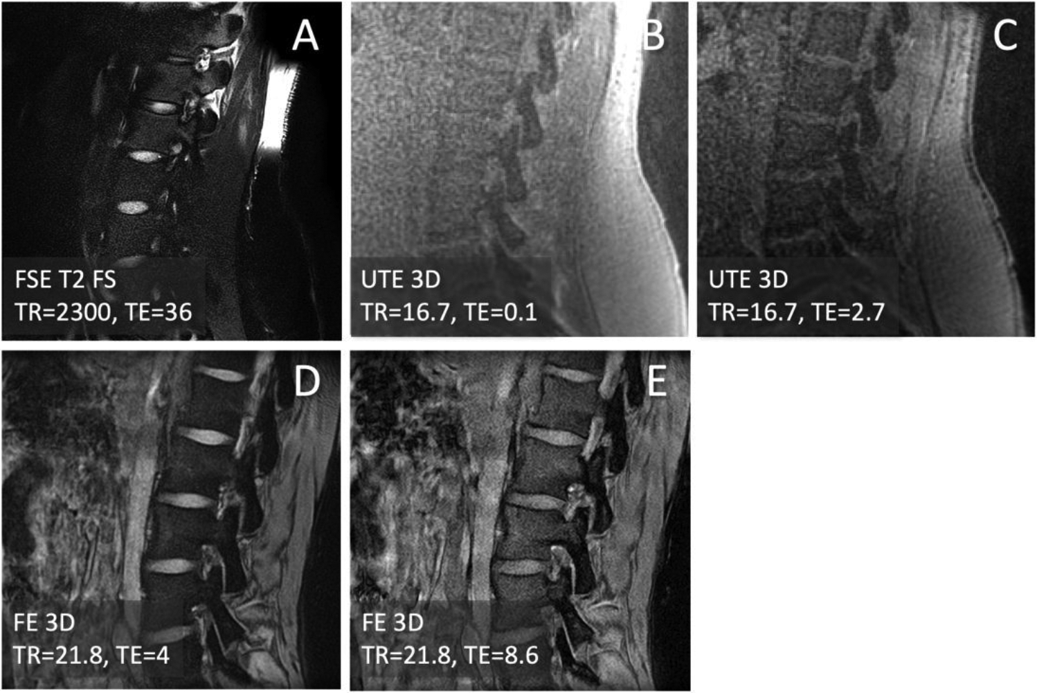
Isthmic spondylolysis is characterized by a stress injury to the pars interarticularis bones of the lumbar spines, often missed by conventional magnetic resonance imaging (MRI) necessitating a computed tomography (CT) for accurate diagnosis. We compare MRI techniques suitable for producing CT-like images. Lumbar spines of asymptomatic and low back pain (LBP) subjects were imaged at 3-Tesla with multi-echo ultrashort echo time (UTE) and field echo (FE) sequences followed by simple post-processing of averaging and inverting to depict spinal bone with CT-like appearance. Contrast-to-noise ratio (CNR) for bone was determined to compare UTE vs. FE and single-echo vs. multi-echo images. Visually, both sequences depicted cortical bone with good contrast; UTE-processed provided a flatter contrast for soft tissues that made it easy to distinguish from bone, while FE-processed images had better resolution and bone-muscle contrast, important for fracture detection. Additionally, multiecho images provided significantly (p=0.03) greater CNR compared single-echo. Using these techniques, a progressive spondylolysis was detected in a LBP subject. This study has demonstrated the feasibility of spine bone MRI to yield CT-like contrast. Through the employment of multiecho UTE and FE sequences combined with simple processing, we have observed enhancements in image quality and contrast, sufficient to detect pars fracture.
Keywords: Bone Fracture; Low Back Pain; MRI; Pars Interarticularis.
Conflict of interest statement
Conflicts of Interest: Mr. Yamashita is an employee of Canon Medical Systems, Japan. Remaining authors declare no conflicts of interest.
Figures


Similar articles
-
Fast Volumetric Imaging of Bone Using a Three-Dimensional Short TR Adiabatic Inversion Recovery Ultrashort Echo Time (STAIR-UTE) Sequence.NMR Biomed. 2025 Sep;38(9):e70102. doi: 10.1002/nbm.70102. NMR Biomed. 2025. PMID: 40664492
-
Deep-Learning-Aided Evaluation of Spondylolysis Imaged with Ultrashort Echo Time Magnetic Resonance Imaging.Sensors (Basel). 2023 Sep 21;23(18):8001. doi: 10.3390/s23188001. Sensors (Basel). 2023. PMID: 37766055 Free PMC article.
-
A comprehensive set of ultrashort echo time magnetic resonance imaging biomarkers to assess cortical bone health: A feasibility study at clinical field strength.Bone. 2024 Apr;181:117031. doi: 10.1016/j.bone.2024.117031. Epub 2024 Feb 2. Bone. 2024. PMID: 38311304 Free PMC article.
-
Computed tomography versus magnetic resonance imaging versus bone scintigraphy for clinically suspected scaphoid fractures in patients with negative plain radiographs.Cochrane Database Syst Rev. 2015 Jun 5;2015(6):CD010023. doi: 10.1002/14651858.CD010023.pub2. Cochrane Database Syst Rev. 2015. PMID: 26045406 Free PMC article.
-
The Black Book of Psychotropic Dosing and Monitoring.Psychopharmacol Bull. 2024 Jul 8;54(3):8-59. Psychopharmacol Bull. 2024. PMID: 38993656 Free PMC article. Review.
References
-
- Wiltse LL; Newman PH; Macnab I Classification of spondylolisis and spondylolisthesis. Clin Orthop Relat Res 1976, 23–29. - PubMed
-
- Fredrickson BE; Baker D; McHolick WJ; Yuan HA; Lubicky JP The natural history of spondylolysis and spondylolisthesis. J Bone Joint Surg Am 1984, 66, 699–707. - PubMed
-
- Bechtel W; Griffiths H; Eisenstadt R The Pathogenesis of Spondylolysis. Investigative radiology 1982, 17, S29.
Grants and funding
LinkOut - more resources
Full Text Sources
Miscellaneous