. 2025 May 20:12:rbaf046.
doi: 10.1093/rb/rbaf046. eCollection 2025.

Construction of EGCG/chlorhexidine functionalized coating to reinforce the soft tissue seal at transmucosal region of implants

Affiliations

Construction of EGCG/chlorhexidine functionalized coating to reinforce the soft tissue seal at transmucosal region of implants

Lijie Zhang et al. Regen Biomater. .

Abstract

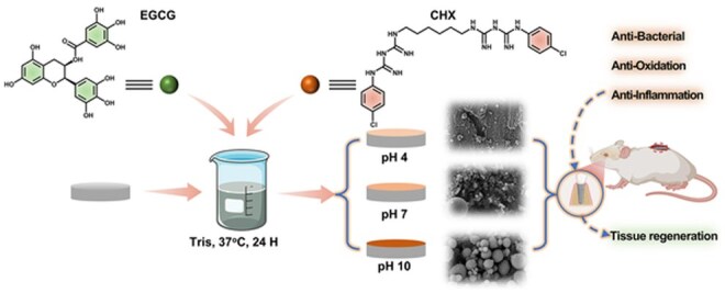
Recent advancements in dental implant technology have provided more reliable and durable solutions for patients. Soft tissue seal (STS) is crucial for achieving implant stability, maintaining tissue health and promoting integration with surrounding soft and hard tissues. However, the STS around implants is fragile and susceptible to disruption by oral pathogens, particularly in patients with periodontitis or poor oral hygiene, leading to complications such as peri-implant mucositis and peri-implantitis. To promote STS formation, it is crucial to maintain the balance between bacterial and host cells while effectively managing inflammation. Although titanium-based implants exhibit biocompatibility, they lack inherent antibacterial and anti-inflammatory properties. To address these challenges, we developed a dual-function antibacterial and anti-inflammatory coating using chlorhexidine (CHX) and epigallocatechin gallate (EGCG). CHX effectively reduces bacterial adhesion but may inhibit fibroblast proliferation, while EGCG provides antioxidant and anti-inflammatory benefits. Three types of EGCG/CHX composite coatings were developed on titanium surfaces at different pH values. These coatings exhibited enhanced bacterial resistance, reduced inflammation and ROS scavenging capabilities, with higher pH levels further improving their performance. In vivo studies also confirmed that these coatings effectively prevented bacterial adhesion, mitigated inflammation and promoted STS formation, thereby holding significant promise for enhancing the long-term success of dental implants.

Keywords: STS; anti-inflammatory; antibacterial; dental implant.


Figures

None
Graphical abstract
Figure 1.
Figure 1.
(A) SEM images of the surfaces of samples from different groups; (B) AFM characterization of coating morphology on titanium sheet surfaces; (C) the surface roughness of the sample; (D) wide-scan XPS spectra; (E) atomic percentage analysis; (F) O1s peak fittings of different pH values; (G) FTIR analysis; (H) the release curve of EGCG; (I) the release curve of CHX.
Figure 2.
Figure 2.
Antibacterial effect on the surface of sample before release. (A) SEM observation of surface bacteria on samples with different components; (B) liquid antibacterial bacterial plate culture results; (C) and counting results; (D) different groups of samples antibacterial zones; (E) staining of live/dead bacteria results.
Figure 3.
Figure 3.
Antibacterial effect on the surface of sample after release. (A) SEM observation of surface bacteria on samples with different components; (B) staining of live/dead bacteria results; (C) liquid antibacterial bacterial plate culture counting results of samples after release.
Figure 4.
Figure 4.
Adhesion and proliferation of fibroblasts on different groups of sample surfaces. (A) Fluorescence staining results; (B) CCK-8 results; (C) bacterial/cell co-culture (L929/S. a) on sample surface; (D) fluorescence staining results of surface cells after sample release; (E) bacterial/cell co-culture on surface after sample release. (F) CCK-8 results of cells after sample release.
Figure 5.
Figure 5.
The antioxidant effect on the surface of samples from different groups. (A) EGCG mechanism for scavenging free radicals; (B) the effect of samples on scavenging DPPH free radicals; (C) and scavenging ABTS+ free radicals; (D) RT-qPCR and Elisa determination of pro-inflammatory cytokine levels; (E) flow cytometry data of macrophages; (F) fluorescence staining results of fibroblasts; (G) data of flow cytometry for detection of reactive oxygen species in macrophages 7 days after sample release; (H) data of flow cytometry for detection of reactive oxygen species in macrophages 14 days after sample release; (I) and fluorescence staining results of fibroblasts after sample release.
Figure 6.
Figure 6.
In vivo evaluation of back implants in rats. (A) Subcutaneous implantation model in rats to evaluate in vivo antibacterial and anti-inflammatory properties: modified materials were implanted subcutaneously followed by bacterial injection to simulate clinical infection; implants were harvested on days 1 and 7 for bacterial quantification using contact and spread plate methods, and tissue samples were collected at 1 month for histological analysis. (B) Press plate and spread-plate results of rat subcutaneous implants after removal 1 day and 7 days. (C) HE staining of the fibrous capsule surrounding the materials. (D) Thickness measurement of the fibrous capsule. (E) The proportion of inflammatory cells in the fibrous capsule.
Figure 7.
Figure 7.
In vivo evaluation of dental implants in rats. (A) Implantation of dental implants in rats and the process of their performance evaluation; (B) HE staining of the periodontal tissues surrounding the implants in rats(“★”is the implant placement position); (C) quantitative analysis of inflammatory factors in the periodontal tissues surrounding the implants in rats.
Figure 8.
Figure 8.
Scheme of design of EGCG/CHX coating to manage inflammation through antibacterial and antioxidation to boost STS though cell adherence promotion for the prevention and treatment of peri-implantitis.

Similar articles

References

    1. Jin S, Yu Y, Zhang T, Xie D, Zheng Y, Wang C, Liu Y, Xia D. Surface modification strategies to reinforce the soft tissue seal at transmucosal region of dental implants. Bioact Mater 2024;42:404–32. - PMC - PubMed
    1. Alves CH, Russi KL, Rocha NC, Bastos F, Darrieux M, Parisotto TM, Girardello R. Host-microbiome interactions regarding peri-implantitis and dental implant loss. J Transl Med 2022;20:425. - PMC - PubMed
    1. Wang S, Yang Y, Li W, Wu Z, Li J, Xu K, Zhang W, Zheng X, Chen J. Study of the relationship between chlorhexidine-grafted amount and biological performances of micro/nanoporous titanium surfaces. ACS Omega 2019;4:18370–80. - PMC - PubMed
    1. Jiang X, Yao Y, Tang W, Han D, Zhang L, Zhao K, Wang S, Meng Y. Design of dental implants at materials level: an overview. J Biomed Mater Res A 2020;108:1634–61. - PubMed
    1. Zhou N, Dong H, Zhu Y, Liu H, Zhou N, Mou Y. Analysis of implant loss risk factors especially in maxillary molar location: a retrospective study of 6977 implants in Chinese individuals. Clin Implant Dent Relat Res 2019;21:138–44. - PubMed

LinkOut - more resources