

Recent advances in deep learning for lymphoma segmentation: Clinical applications and challenges
- PMID: 40735544
- PMCID: PMC12304644
- DOI: 10.1177/20552076251362508
Recent advances in deep learning for lymphoma segmentation: Clinical applications and challenges
Abstract
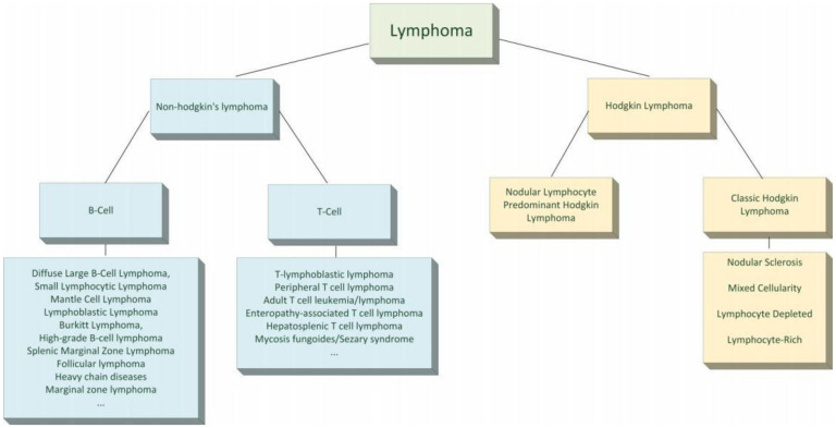
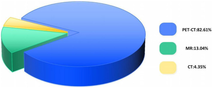
Lymphoma is a prevalent malignant tumor within the hematological system, posing significant challenges to clinical practice due to its diverse subtypes, intricate radiological and metabolic manifestations. Lymphoma segmentation studies based on positron emission tomography/computed tomography (PET/CT), CT, and magnetic resonance imaging represent key strategies for addressing these challenges. This article reviews the advancements in lymphoma segmentation research utilizing deep learning methods, offering a comparative analysis with traditional approaches, and conducting an in-depth examination and summary of aspects such as dataset characteristics, backbone networks of models, adjustments to network structures based on research objectives, and model performance. The article also explores the potential and challenges of translating deep learning-based lymphoma segmentation research into clinical scenarios, with a focus on practical clinical applications. The future research priorities in lymphoma segmentation are identified as enhancing the models' clinical generalizability, integrating into clinical workflows, reducing computational demands, and expanding high-quality datasets. These efforts aim to facilitate the broad application of deep learning in the diagnosis and treatment monitoring of lymphoma.
Keywords: Lymphoma; artificial intelligence; clinical application; deep learning; medical image segmentation.
© The Author(s) 2025.
Conflict of interest statement
The authors declared no potential conflicts of interest with respect to the research, authorship, and/or publication of this article.
Figures
References
-
- Connors JM, Cozen W, Steidl C, et al. Hodgkin lymphoma. Nat Rev Dis Primers 2020; 6: 61. - PubMed
-
- Lu P. Staging and classification of lymphoma. Semin Nucl Med 2005; 35: 160–164. Elsevier. - PubMed
-
- Lewis RB, Mehrotra AK, Rodríguez P, et al. From the radiologic pathology archives: gastrointestinal lymphoma: radiologic and pathologic findings. Radiographics 2014; 34: 1934–1953. - PubMed
-
- Ramesh KKD, Kiran Kumar G, Swapna K, et al. A review of medical image segmentation algorithms. EAI Endorsed Transact Pervasive Health Technol 2021; 7: e6–e6.
Publication types
LinkOut - more resources
Full Text Sources
