

doi: 10.1016/j.eurox.2025.100414.
eCollection 2025 Sep.
Clinical guidance VVOG: Antenatal care for twin pregnancies
Affiliations
- PMID: 40735655
- PMCID: PMC12304725
- DOI: 10.1016/j.eurox.2025.100414
Item in Clipboard
Clinical guidance VVOG: Antenatal care for twin pregnancies
Eur J Obstet Gynecol Reprod Biol X.
.
No abstract available
Conflict of interest statement
The authors declare that they have no known competing financial interests or personal relationships that could have appeared to influence the work reported in this paper.
Figures
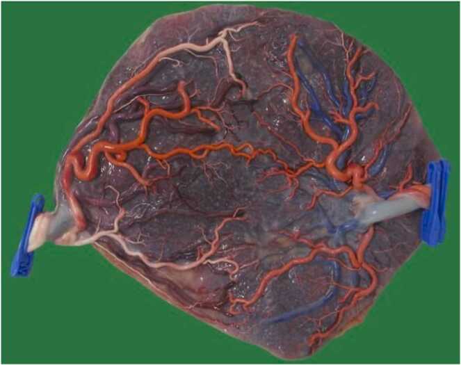
Placenta of uncomplicated monochorionic diamniotic twins born at 36 weeks after dye injection demonstrating the different blood vessel connections. The amnion and intertwinseptum were removed

Ultrasound image of dichorionic twins at 12 weeks. The intertwinseptum consists of a thick chorionic layer with two thin amniotic membranes (amnion-chorion-amnion) along both sides. The chorionic layer inserts at the level of the placenta as a "full lambda" sign
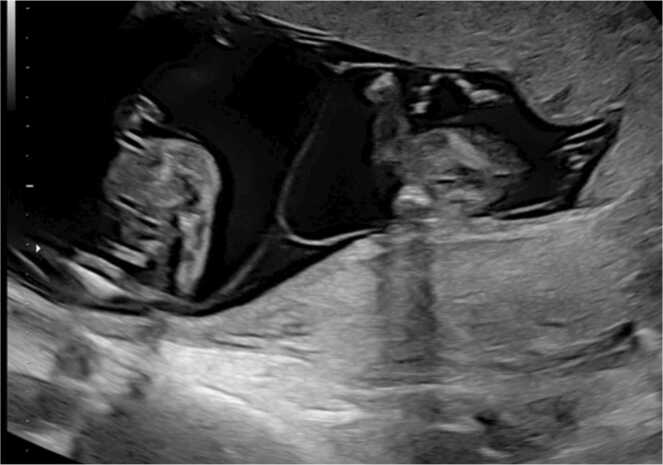
Image of monochorionic diamniotic twins at 13 weeks. The intertwinseptum consists only of two thin amniotic membranes (amnion-amnion) that inserts as an "empty lambda" sign

Image of dichorionic twins at 9 weeks. There are two separate gestational sacs and each fetus and yolk sac is in a separate exocoelomic cavity

Ultrasound image of monochorionic twins at 10 weeks. There is one gestational sac and a common exocoelomic cavity containing both fetuses and yolk sacs

Ultrasound image of monoamniotic twins at 13 weeks. Both fetuses are in a common amniotic sac with umbilical cords already visibly entangled

Schematic for the ultrasound follow-up of di- and monochorionic twins

Twin-to-twin transfusion syndrome is characterized by a severe difference in amniotic fluid with a deepest vertical amniotic fluid loge less than 2 cm in the donor who has a small or empty bladder and more than 8 cm (before 20 weeks) or more than 10 cm (after 20 weeks) in the receptor who has a distended bladder
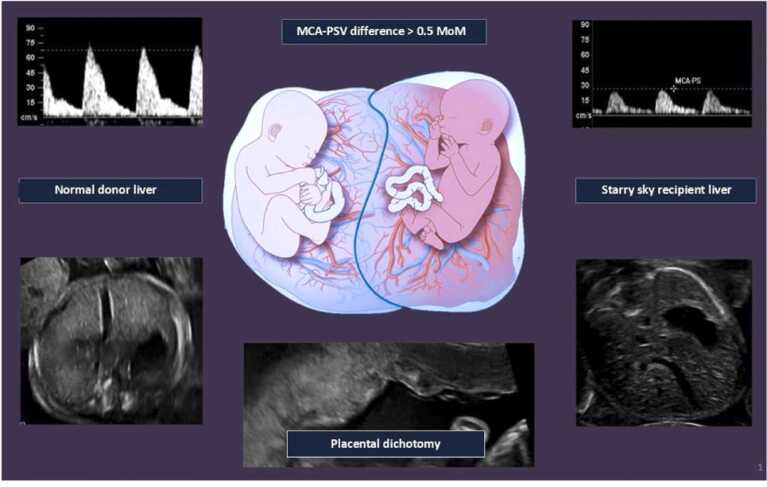
Twin anemia polycytemia sequence is characterized by a severe difference in hemoglobin with an increased peak velocity in the middle cerebral artery (MCA PSV) of 1.5 MoM (multiples of the median) or more in the anemic donor and 0.8 MoM or less in the polycytemic receptor. A difference of 1 MoM or more is also considered diagnostic. Often there is also an echogenicity difference of the placenta and the receptor has a dark congested liver in which the small venules light up ("starry sky")
References
-
- Béatrice Blondel M.K. Trends in the occurrence, determinants, and consequences of multiple births. Semin Perinatol. 2002;26(4):239–249. - PubMed
-
- Devlieger R., Goemaes R., Laubach M. Perinatal health in Flanders - Year 2020. Bruss Study Cent Perinat Epidemiol. 2021
-
- Santana D.S., Cecatti J.G., Surita F.G., Silveira C., Costa M.L., Souza J.P., et al. Twin Pregnancy and severe maternal outcomes: the world health organization multicountry survey on maternal and newborn health. Obstet Gynecol. 2016;127(4):631–641. - PubMed
-
- National Institution for Health and Care Excellence. Twin and triplet pregnancy NICE guideline (2019).
-
- Society of Obstetrician and Gynaecologists of Canada SOGC clinical practice guidance: Management of Dichorionic Twin Pregnancies. J Obstet Gynecol Can. 2022;44(7):819–834. - PubMed
LinkOut - more resources
Full Text Sources
