Review
. 2025 Jul 13:34:102066.
doi: 10.1016/j.mtbio.2025.102066. eCollection 2025 Oct.

Topography-based implants for bone regeneration: Design, biological mechanism, and therapeutics

Affiliations
Review

Topography-based implants for bone regeneration: Design, biological mechanism, and therapeutics

Weiwei Guo et al. Mater Today Bio. .

Abstract

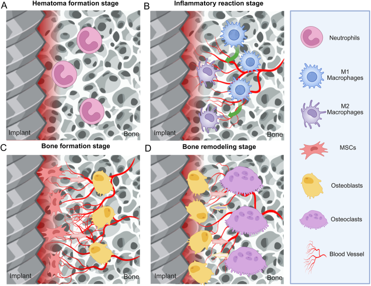
With the increasing demand for bone defect repair, bone implant materials have emerged as a critical alternative to traditional autologous or allogeneic bone grafts. However, their clinical performance remains limited due to challenges such as prolonged healing times and suboptimal repair quality. Moreover, in patients with certain pathological conditions (e.g., diabetes mellitus and osteoporosis), disruptions in the bone microenvironment further compromise regenerative outcomes. To address these limitations, surface modification strategies have been developed to regulate implant-bone tissue interactions and improve therapeutic efficacy. This review systematically summarizes recent advances in bone regeneration implants with a focus on topographical modifications, encompassing design principles, underlying biological mechanisms, and therapeutic applications. Particular attention is given to the influence of implant surface topography on the biological behaviors of osteoblasts, osteoclasts, and macrophages within the bone microenvironment, as well as their responses under complex pathological and physiological conditions. The review also discusses current challenges related to achieving micro/nanoscale structural balance, personalization, and clinical translation of implant surface topographies, and highlights future directions in precision bone regeneration through multidisciplinary approaches, artificial intelligence-driven optimization, and long-term clinical validation. Collectively, these insights may inform future research on bone implant materials and support the development of novel strategies for personalized treatment of bone defect repair.

Keywords: Biological mechanisms; Bone regeneration; Implants; Surface morphology; Translational applications.


Conflict of interest statement

The authors declare that they have no known competing financial interests or personal relationships that could have appeared to influence the work reported in this paper.

Figures

Image 1
Graphical abstract
Fig. 1
Fig. 1
Schematic illustration of implant-bone microenvironment interactions regulating bone repair processes. A) Hematoma formation stage. B) Inflammatory reaction stage. C) Bone formation stage. D) Bone remodeling stage. Created in BioRender. Abbreviations: MSCs: Mesenchymal stem cells.
Fig. 2
Fig. 2
Surface morphology of bone implants prepared by physical and chemical methods. (A) Fabrication and applications of surface micro/nanostructures by femtosecond laser. a. Schematic diagram. b. SEM images with different pulse energies. c. SEM images after femtosecond laser treatment and photographs of liquid metal patterns of different shapes [148]. (B) Micro/nano-mimetic morphology preparation and promotion of bone integration in rabbits. a. Morphology of BMSCs on bionic surfaces. b. Osteogenic gene expression. c. Qualitative images of mineralization formation and collagen type I. d. 3D Micro-CT images and quantitative results of BIC [172]. Abbreviations: LIPSS: Laser-induced periodic surface structures; SEM: Scanning electron microscope; BMSCs: Mesenchymal stem cells; Micro-CT: Micro-computed tomography; BIC: Bone-to-implant contact.
Fig. 3
Fig. 3
Mechanisms of osteoblast signaling pathways in the implant-bone microenvironment. Created in BioRender. Abbreviations: LRP: Low-density lipoprotein receptor-related protein; FZD: Frizzled; APC: Adenomatous polyposis coli; PLC: Phospholipase C; PKC: Protein kinase C; TAK1: TGF-β activated kinase 1; CAMKII: Calcium/calmodulin-dependent protein kinase II; NFAT: Nuclear factor of activated t-cells; DAAM: Dvl-associated activator of morphogenesis 1; RAC1: Ras-related C3 botulinum toxin substrate 1; JNK: c-Jun N-terminal kinase; ROCK: Rho-associated protein kinase; PCP: Planar cell polarity; BMP: Bone morphogenetic proteins; SMAD: Small mother against decapentaplegic; FAK: Focal adhesion kinase; TGF-β: Transforming growth factor-β; TCF: T-cell factor; LEF: Lymphoid enhancer-binding factor; RUNX2: Runt-related transcription factor 2; OPN: Osteopontin; OCN: Osteocalcin; ALP: Alkaline phosphatase; COL1: Collagen type 1.
Fig. 4
Fig. 4
Mechanisms of osteoclast signaling pathways in the implant-bone microenvironment. Created in BioRender. Abbreviations: FAK: Focal adhesion kinase; JNK: c-Jun N-terminal kinase; RANK: Receptor activator of nuclear factor kappa-B; RANKL: Receptor activator of nuclear factor kappa-B ligand; TRAF6: TNF receptor-associated factor 6; MAPK: Mitogen-activated protein kinase; M-CSF: Macrophage colony-stimulating factor; CSF-1R: Colony stimulating factor 1 receptor; PI3K: Phosphoinositol-3 kinase; Akt: Protein kinase B; TRAP: Tartrate-resistant acid phosphatase; CTSK: Cathepsin K; MMP9: Matrix metalloproteinase 9; NFATc1: Nuclear factor of activated T-cells, cytoplasmic, calcineurin-dependent 1.
Fig. 5
Fig. 5
Mechanisms of macrophage signaling pathways in the implant-bone microenvironment. Created in BioRender. Abbreviations: MHC-II: Major histocompatibility complex, class II; TLR: Toll-like receptor; CD86: Cluster of differentiation 86; FIZZ1: Fibrinogen-like protein 1; Ym1/2: Macrophage inflammatory protein 3, neutrophil gelatinase-associated lipocalin; FAK: Focal adhesion kinase; SMAD: Small mother against decapentaplegic; PI3K: Phosphoinositol-3 kinase; Akt: Protein kinase B; MyD88: Myeloid differentiation primary response 88; STAT: Signal transducer and activator of transcription; IRF: Interferon regulatory factors; KLF-4: Krüppel-like factor 4; IL-10: Interleukin 10; BMP-2: Bone morphogenetic protein 2; VEGF: Vascular Endothelial Growth Factor; IGF-1: Insulin-like Growth Factor 1; CCL-18: Chemokine (C-C Motif) Ligand 18.
Fig. 6
Fig. 6
Topology-based implants for in vivo treatment of animal models of critical bone defects. (A) Nanoscale and hybrid micro- and nanocomposite surfaces for intraosseous implants. a. SEM images. b. Confocal images of HMSCs. c. Ground sections of bone-implant interface. d. Bone micromorphometry parameters [469]. (B) Natural micropatterned fish scales combing direct osteogenesis and osteoimmunomodulatory functions for enhancing bone regeneration. a. Schematic illustration. b. SEM images. c. Critical bone defect modeling and implant filling. d. Micro-CT images. e. Quantitative micro-CT images analysis [492]. Abbreviations: SEM: Scanning electron microscope; HMSCs: Human mesenchymal stem cells; Micro-CT: Micro-computed tomography.
Fig. 7
Fig. 7
Topology-based implants for in vivo treatment of dental implants. (A) Nested nanofiber structure of titanium nano bowls. a. Preparation schematic. b. EDS mapping images and surface roughness. c. 2D and 3D AFM images. d. FE-SEM images e. SEM image of BMSCs at 24 h [494]. (B) Nanofiber structure promotes osteointegration of Beagle dog Implants. a. Schematic illustration. b. XRD patterns and XPS patterns. c. Micro CT images. d. BV/TV, Tb.Th, BS/BV and BMD [494]. Abbreviations: TNT: TiO2 nanotubes; TNB: TiO2 nanobowl; NTNF: Nest-like titanite nanofiber structure; AFM: Atomic force microscope; EDS: Energy dispersive spectroscopy; FE-SEM: Field-emission scanning electron microscope; XRD: X-ray diffraction; XPS: X-ray photoelectron spectroscopy; BV/TV: Bone volume/total volume; Tb.Th: Trabecular thickness; BS/BV: Bone surface/bone volume; BMD: Bone mineral density.
Fig. 8
Fig. 8
Topology-based implants for in vivo treatment of diabetic bone defects. (A) M2 macrophage-derived exosomes promote diabetic fracture healing by acting as an immunomodulator. a. Localization of M1 macrophages. b. Macrophage mRNA expression levels. c. FCM analysis. d. Micro-CT images. e. SOFG staining of healing tissue sections [507]. (B) Re-establishment of macrophage homeostasis by titanium surface modification in type II diabetes promotes osseous healing. a. Diagram of the molecular mechanism. b. Cytokine secretion profile of M1-polarised GK macrophages. c. Immunofluorescence images. d. Toluidine blue-stained histologic sections [65]. Abbreviations: M0: Unstimulated macrophage; Pre-Obs: Pre-osteoblasts; Obs: Osteoblasts; GK: Goto kakizaki; SLA: Sandblast; modSLA: Modified hydrophilic sandblast; FCM: Flow cytometry; Micro-CT: Micro-computed tomography; SOFG: Standards and ontologies for functional genomics.
Fig. 9
Fig. 9
Topology-based implants for in vivo treatment of osteoporotic bone defects. (A) Liver-inspired polyetherketoneketone scaffolds. a. Schematic illustration. b. Live/dead fluorescence, SEM images and cytoskeleton of RAW264.7 cells [495]. (B) Polyetherketoneketone Scaffolds Promote Osteoporotic Osteointegration. a. Schematic illustration. b. SEM images. c. 3D surface optical profiles. d. Expression of osteogenic genes in BMSCs. f. 3D images of micro-CT, quantitative analysis of bone density and bone volume fraction, and Van Gieson staining [495]. Abbreviations: SEM: Scanning electron microscope; PEKK: Polyetherketoneketone; PEKK‐L: Sulfonating PEKK with 80 % H2SO4; PEKK‐SW: Sulfonating PEKK with 98 % H2SO4.
Fig. 10
Fig. 10
Future prospects for topography-based bone implants. Created in BioRender. Abbreviations: BMP-2: Bone morphogenetic protein 2; VEGF: Vascular endothelial growth factor; TNF-α: Tumor necrosis factor alpha; IL-1β: Interleukin 1 beta; TGF-β: Transforming growth factor beta; IL-10: Interleukin 10.

Similar articles

References

    1. Liang W., Zhou C., Bai J., Zhang H., Long H., Jiang B., Dai H., Wang J., Zhang H., Zhao J. Current developments and future perspectives of nanotechnology in orthopedic implants: an updated review. Front. Bioeng. Biotechnol. 2024;12 doi: 10.3389/fbioe.2024.1342340. - DOI - PMC - PubMed
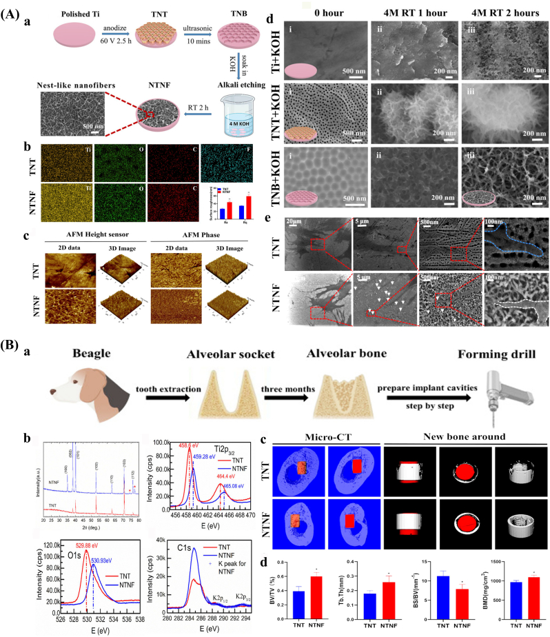
    1. Li W., Su Z., Hu Y., Meng L., Zhu F., Xie B., Zhou Z., Cui S., Wang M., Wu Q., Yao S. Functional and structural construction of photothermal-responsive PEEK composite implants to promote bone regeneration and bone-implant integration. Compos. Sci. Technol. 2024;258 doi: 10.1016/j.compscitech.2024.110885. - DOI
    1. Li J., Ma J., Feng Q., Xie E., Meng Q., Shu W., Wu J., Bian L., Han F., Li B. Building osteogenic microenvironments with a double-network composite hydrogel for bone repair. Research. 2023;6:21. doi: 10.34133/research.0021. - DOI - PMC - PubMed
    1. Brochu B.M., Sturm S.R., Kawase D.Q.G.J., Mirsky N.A., Sandino A.I., Panthaki K.Z., Panthaki K.Z., Nayak V.V., Daunert S., Witek L., Coelho P.G. Advances in bioceramics for bone regeneration: a narrative review. Biomimetics (Basel). 2024;9(11) doi: 10.3390/biomimetics9110690. - DOI - PMC - PubMed
    1. Baldwin P., Li D.J., Auston D.A., Mir H.S., Yoon R.S., Koval K.J. Autograft, allograft, and bone graft substitutes: clinical evidence and indications for use in the setting of orthopaedic trauma surgery. J. Orthop. Trauma. 2019;33(4):203–213. doi: 10.1097/BOT.0000000000001420. - DOI - PubMed

LinkOut - more resources