

doi: 10.1002/ccr3.70686.
eCollection 2025 Aug.
A Typical Dermoscopic Pattern of PLEVA
Affiliations
- PMID: 40735706
- PMCID: PMC12301154
- DOI: 10.1002/ccr3.70686
Item in Clipboard
A Typical Dermoscopic Pattern of PLEVA
Clin Case Rep.
.
Abstract
PLEVA is a benign inflammatory disorder. Dermoscopy is a non-invasive diagnostic modality. Thus, the new dermoscopic pattern mentioned in our literature can aid in the diagnosis of the disease, unlike the skin biopsy, which is an invasive modality and shows non-specific features in PLEVA.
Keywords: PLEVA; central crust; dermoscopy; targetoid pattern; vascular structures; white scales.
© 2025 The Author(s). Clinical Case Reports published by John Wiley & Sons Ltd.
Conflict of interest statement
The authors declare no conflicts of interest.
Figures
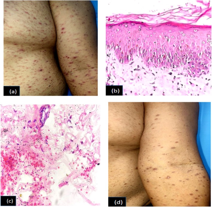
(a) PLEVA: Mulitple erythematous macules, papules, crusted lesions with mica like scales over trunk and extremities, 1 (b) Hematoxylin and eosin (H/E) stain (40×): Parakeratosis, spongiosis and vacuolar degeneration in basal layer of epidermis, 1 (c) H/E stain (40×): Extravasation of RBCs in dermis, 1 (d) Multiple hyperpigmented macules and papules following 3 weeks of treatment with doxycycline.
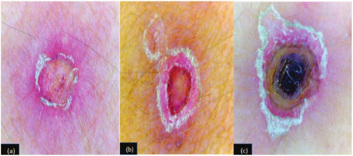
(a) Dermoscopy PLEVA: Central crusting, reddish areas with polymorphic vessels surrounded by peripheral white scales, 2 (b) Typical target pattern of central crust, intermediate vascular ring surrounded by white scales on pinkish background, 2 (c) Central necrotic areas around the hair follicles with peripheral crusting and scaling.
Similar articles
-
Pityriasis lichenoides et varioliformis acuta in skin of color: new observations by dermoscopy.Dermatol Pract Concept. 2017 Jan 31;7(1):27-34. doi: 10.5826/dpc.0701a05. eCollection 2017 Jan. Dermatol Pract Concept. 2017. PMID: 28243491 Free PMC article.
-
Line-field confocal optical coherence tomography of basal cell carcinoma: A retrospective study on diagnostic performance.J Eur Acad Dermatol Venereol. 2025 Aug;39(8):1468-1480. doi: 10.1111/jdv.20459. Epub 2024 Dec 2. J Eur Acad Dermatol Venereol. 2025. PMID: 39620273
-
Dermoscopy of cutaneous adnexal tumours: a systematic review of the literature.J Eur Acad Dermatol Venereol. 2022 Sep;36(9):1524-1540. doi: 10.1111/jdv.18210. Epub 2022 May 25. J Eur Acad Dermatol Venereol. 2022. PMID: 35536546 Free PMC article.
-
Skin-CAD: Explainable deep learning classification of skin cancer from dermoscopic images by feature selection of dual high-level CNNs features and transfer learning.Comput Biol Med. 2024 Aug;178:108798. doi: 10.1016/j.compbiomed.2024.108798. Epub 2024 Jun 25. Comput Biol Med. 2024. PMID: 38925085
-
Changes in melanocytic nevi treated with laser hair removal: A systematic review.Lasers Surg Med. 2023 Sep;55(7):617-624. doi: 10.1002/lsm.23712. Epub 2023 Jul 26. Lasers Surg Med. 2023. PMID: 37493510
References
-
- Teklehaimanot F., Gade A., and Rubenstein R., “Pityriasis Lichenoides et Varioliformis Acuta (PLEVA),” in StatPearls (StatPearls Publishing, 2024). - PubMed
-
- Bhut A., Shah A., and Nair P. A., “Dermatoscopic Findings of Pityriasis Lichenoides et Varioliformis Acuta,” Indian Journal of Paediatric Dermatology 21 (2020): 249–250.
-
- Virdi S. K., Kanwar A. J., and Saikia U. N., “Pityriasis Lichenoides With Ulceronecrosis and Hyperthermia: A Rare Variant of Pityriasis Lichenoides et Varioliformis Acuta,” Indian Journal of Dermatology, Venereology and Leprology 76 (2010): 172–175. - PubMed
LinkOut - more resources
Full Text Sources
