. 2025 Aug;603(16):4637-4657.
doi: 10.1113/JP287804. Epub 2025 Jul 30.

Transcutaneous spinal random noise stimulation enhances motor memory consolidation and corticospinal transmission in humans

Affiliations

Transcutaneous spinal random noise stimulation enhances motor memory consolidation and corticospinal transmission in humans

Mitsuhiro Nito et al. J Physiol. 2025 Aug.

Abstract

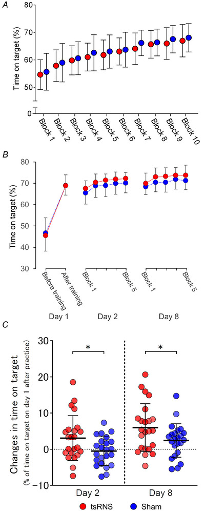
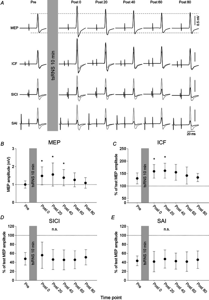
Stochastic resonance sensory input modulates the central nervous system's excitability, thereby possibly influencing motor skill learning and retention. We investigated the effects of transcutaneous spinal random noise stimulation (tsRNS) at the cervical level on motor skill learning and corticospinal transmission in healthy humans. Participants performed a 20 min visuomotor tracking training task requiring rapid shifts in pinch force, with motor performance tests conducted before, immediately after, 1 day after and 7 days after the training to assess motor skill learning and retention. During the task, participants received real or sham tsRNS for 20 and 0.5 min, respectively. Motor performance improved equally in both groups immediately after training; however, the real tsRNS group showed a higher performance than the sham group at 1 and 7 days post-training. Beta-band corticomuscular coherence increased immediately after training in both groups, and higher performance on 1 day after the training was positively correlated with a greater change in corticomuscular coherence. To elucidate the mechanisms contributing to the enhanced motor memory retention induced by tsRNS, we investigated its effects on cortical and spinal excitability. We observed increased intracortical facilitation and somatosensory evoked potential amplitude following tsRNS; however, the efficacy of cortico-motoneuronal synaptic transmissions and the excitability of spinal motoneurons remained unchanged. Collectively, tsRNS can enhance the corticospinal drive to spinal motoneurons indirectly by increasing the ascending afferent input strength and cortical excitability via the augmented activity of facilitatory interneurons, resulting in improved motor memory retention. Thus, tsRNS may have important clinical applications for rehabilitation after central nervous system lesions. KEY POINTS: Stochastic resonance sensory input modulates the excitability of the central nervous system and may influence motor skill learning and motor memory retention. Transcutaneous spinal random noise stimulation (tsRNS) applied at the cervical level can enhance motor skill learning and motor memory retention in healthy humans. tsRNS can increase the ascending afferent input to the cortex and the excitability of the intracortical circuits rather than directly modulating the descending motor output, resulting in improved motor memory retention. These findings suggest that tsRNS is an effective strategy for promoting functional motor recovery of the upper limb after the development of central nervous system lesions.

Keywords: motor skill learning; precision grip; rehabilitation; spinal stimulation; transcranial magnetic stimulation; upper limb.


Conflict of interest statement

The authors declare that they have no competing interests.

Figures

Figure 1
Figure 1. Experimental setup and protocol
A, transcutaneous spinal random noise stimulation (tsRNS) was delivered through saline‐soaked sponge electrodes placed on the midline of the anterior neck and over the spinous process of the seventh cervical vertebra. B, motor performance assessment. Participants controlled the vertical position of a cursor by adjusting the pinch force. Blue line represents the cursor trajectory. Performance was scored as the proportion (%) of time the cursor remained within the target boxes. C, time course of Experiment 2. MVC, maximum voluntary contraction. [Colour figure can be viewed at wileyonlinelibrary.com]
Figure 2
Figure 2. Effects of transcutaneous spinal random noise stimulation (tsRNS) at different intensities on motor evoked potentials (MEPs) measured over the first dorsal interosseous (FDI) muscle in response to transcranial magnetic stimulation of the primary motor cortex
A, MEPs measured in the FDI muscle of a single participant before and after tsRNS at the indicated intensities. Each waveform represents the average of 15 trials. B, mean ± SD values obtained from 20 participants. Each symbol indicates the averaged results of 1.0 mA (grey circles), 2.0 mA (white circles) and 3.0 mA tsRNS (black circles). Asterisks indicate significant differences compared with the values before tsRNS (‘Pre’) and the horizontal bars indicate significant differences compared with the other time points (P < 0.05).
Figure 3
Figure 3. Transcutaneous spinal random noise stimulation (tsRNS) during motor training enhances motor memory retention
A, group average on‐target scores during motor training with real tsRNS (red circles) or sham tsRNS (blue circles). B, group average on‐target scores before and after motor training on Day 1 and for the retention tests (blocks 1–5) on day 2 and day 8. C, individual retention rates for blocks 1–5 on day 2 (left) and day 8 (right) are calculated as a percentage of the mean score on day 1 immediately after training. The overlaid black lines show the group average (thick line) with the SD (thin error lines). Each circle in (C) represents individual data points obtained from the real tsRNS group (red, n = 23) and sham tsRNS group (blue, n = 23). [Colour figure can be viewed at wileyonlinelibrary.com]
Figure 4
Figure 4. Influence of transcutaneous spinal random noise stimulation (tsRNS) on corticomuscular coherence
A, representative waveforms of corticomuscular coherence on day 1 before motor training (dashed line) and after motor training (continuous line) obtained from two participants. B, individual changes and group results for peak corticomuscular coherence (real tsRNS group: n = 23; sham tsRNS group: n = 23). [Colour figure can be viewed at wileyonlinelibrary.com]
Figure 5
Figure 5. Changes in corticomuscular coherence after transcutaneous spinal random noise stimulation (tsRNS) are correlated with motor memory retention
Each circle represents individual data point obtained from the real tsRNS group (red) and sham tsRNS group (blue). The abscissa indicates changes in normalised corticomuscular coherence before and after motor training at day 1. The ordinate indicates retention rates for blocks 1–5 on day 2 (A) and day 8 (B), calculated as a percentage of the mean score immediately after training on day 1. The dashed line represents the regression line of all data points. [Colour figure can be viewed at wileyonlinelibrary.com]
Figure 6
Figure 6. Transcutaneous spinal random noise stimulation (tsRNS) enhances motor evoked potentials (MEPs) elicited by transcranial magnetic stimulation (mMEPs) but not transcranial electrical stimulation (eMEPs) in the primary motor cortex
A, MEPs recorded from the first dorsal interosseous muscle of a single participant in response to transcranial magnetic stimulation (black; mMEPs) or transcranial electrical stimulation (red; eMEPs) before, during and after tsRNS. Each waveform represents the average of 15 trials. B and C, time course of mMEPs (B) and eMEPs (C). Regarding mMEPs, the mean ± SD values were obtained from 20 participants in the real tsRNS group and 16 participants in the sham tsRNS group (B). Regarding eMEPs, the mean ± SD values were obtained from 10 participants in both the real and sham tsRNS groups (C). Asterisks indicate significant differences compared with ‘Pre’ (P < 0.05). Dagger indicates that the MEP amplitudes are significantly different between the real and sham tsRNS conditions (P < 0.05). [Colour figure can be viewed at wileyonlinelibrary.com]
Figure 7
Figure 7. Transcutaneous spinal random noise stimulation (tsRNS) does not alter maximal M‐waves (M‐max) or F‐waves
A and B, representative M‐max (A) and F‐waves (B) recorded from the first dorsal interosseous muscle of an individual participant. Black and grey traces represent the average of 30 trials and each single waveform. CE, time courses of M‐max (C), F‐wave persistence (D) and F‐wave amplitudes (E) recorded before, during and after tsRNS. The the mean ± SD values are presented in (C). In (D) and (E), horizontal lines within boxes indicate median and interquartile ranges, whereas whiskers indicate minimum and maximum values. Each datapoint was obtained from 20 participants.
Figure 8
Figure 8. Transcutaneous spinal random noise stimulation (tsRNS) enhances multiple somatosensory evoked potential (SEP) components
A, SEPs elicited by ulnar nerve stimulation at the wrist from a single participant. The waveforms were obtained by averaging 300 trials. B and C, individual changes in each SEP component and group results for real tsRNS (n = 16 for P14/N20, n = 20 for N20/P25 and P25/N33) (B) and sham tsRNS (n = 13 for P14/N20, n = 16 for N20/P25 and P25/N33) (C). Asterisks indicate significant differences between ‘Pre’ and ‘Post 0’ (P < 0.05)
Figure 9
Figure 9. Lasting effects of transcutaneous spinal random noise stimulation (tsRNS) on motor evoked potentials (MEPs) and intracortical facilitation (ICF) but not short‐interval intracortical inhibition (SICI) or short‐latency afferent inhibition (SAI)
A, MEPs recorded from the first dorsal interosseous muscle of a single participant in response to transcranial magnetic stimulation (TMS) of the motor cortex. The top waveforms were obtained by a single TMS at constant intensity. ICF and SICI were obtained by measuring MEPs conditioned by subthreshold TMS and SAI by measuring MEPs conditioned by electrical stimulation of the median nerve at the wrist. Grey and black traces indicate the test and conditioned MEPs, respectively. Each waveform represents the average of 15 trials. BE, time courses of MEP amplitude change in response to single TMS (B), ICF (C), SICI (D) and SAI (E). The means and standard deviations were obtained from 19 participants. Asterisks indicate significant differences compared with ‘Pre’ (P < 0.05).
Figure 10
Figure 10. Effects of transcutaneous spinal random noise stimulation on vital signs
Individual changes and group results for heart rate (A) (n = 13), oxygen saturation level (B) (n = 16) systolic (C) and diastolic blood pressure (D) (n = 10).

Similar articles

References

    1. Abe, T. , Miyaguchi, S. , Otsuru, N. , & Onishi, H. (2019). The effect of transcranial random noise stimulation on corticospinal excitability and motor performance. Neuroscience Letters, 705, 138–142. - PubMed
    1. Allison, T. , McCarthy, G. , Wood, C. C. , Darcey, T. M. , Spencer, D. D. , & Williamson, P. D. (1989). Human cortical potentials evoked by stimulation of the median nerve. I. Cytoarchitectonic areas generating short‐latency activity. Journal of Neurophysiology, 62(3), 694–710. - PubMed
    1. Allison, T. , McCarthy, G. , Wood, C. C. , & Jones, S. J. (1991). Potentials evoked in human and monkey cerebral cortex by stimulation of the median nerve. A review of scalp and intracranial recordings. Brain, 114(6), 2465–2503. - PubMed
    1. Butler, J. E. , & Thomas, C. K. (2003). Effects of sustained stimulation on the excitability of motoneurons innervating paralyzed and control muscles. Journal of Applied Physiology, 94(2), 567–575. - PubMed
    1. Chaieb, L. , Antal, A. , & Paulus, W. (2015). Transcranial random noise stimulation‐induced plasticity is NMDA‐receptor independent but sodium‐channel blocker and benzodiazepines sensitive. Frontiers in Neuroscience, 9. - PMC - PubMed

LinkOut - more resources