. 2025 Sep;116(5):e70042.
doi: 10.1002/bip.70042.

In Vivo Wound Healing and Immune Response Studies of Chitosan Cryogels With Invertebrate Model Organism Galleria mellonella

Affiliations

In Vivo Wound Healing and Immune Response Studies of Chitosan Cryogels With Invertebrate Model Organism Galleria mellonella

Sema Ekici et al. Biopolymers. 2025 Sep.

Abstract

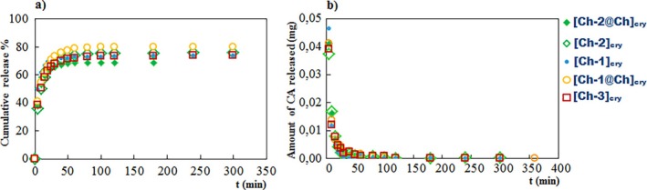
In the present study, it was aimed to prepare single and double network chitosan (Ch) cryogels cross-linked with glutaraldehyde (G), which can be recommended for use as model wound dressings and hemostatic agents, and to reveal in vivo studies with Galleria mellonella. An in vivo study about Ch cryogels with these larvae was not declared in the literature, so our study is the first of its kind. G. mellonella was used to determine the effects of cryogels on immunity, oxidative stress, and wound healing. Cinnamic acid (CA) was loaded onto the cryogels, and the percent cumulative release data of CA were found to be in the range of 69%-80%. The results show that loading of CA onto [Ch-3]cry cryogels considerably improved immune responses; the [Ch-3]cry-CA group was the most successful in terms of immunological response, oxidative stress balance, and wound healing. In accordance with the 3R principles of ethical animal research, the use of G. mellonella in this study served as a scientifically relevant and ethically responsible alternative model to mammals for preliminary assessment of wound healing potential and innate immune activation. The porous structures, high mechanical strengths, and rapidly swelling-deswelling abilities of [Ch-2@Ch]cry and [Ch-3]cry cryogels indicated that these may be suitable for biomedical applications. Analysis of SEM micrographs indicated that the morphology of dual network cryogels prepared in the form of interpenetrating polymeric networks (IPNs) was more regular and homodispersed with respect to single network cryogels. The compressive elasticity modulus (E) values of IPNs cryogels (0.160 N/mm) is approximately 4.6 times that of Ch cryogels with a single network (0.035 N/mm).

Keywords: G. mellonella; chitosan cryogel; full‐IPN cryogel; hemostatic material; wound dressings.


Conflict of interest statement

The authors declare no conflicts of interest.

Figures

SCHEME 1
SCHEME 1
Synthesis procedure of Ch cryogels with single and full‐IPN structure.
FIGURE 1
FIGURE 1
Photos of Ch cryogels after drying with lyophilizer. It is noteworthy that full‐IPN cryogels are larger and have more compact and smooth structure than single cryogels. As the amount of Ch in the cryogels increased, the color of the gels became more yellow due to iminization reaction between Ch and G in weakly acidic conditions.
FIGURE 2
FIGURE 2
Comparative presentation of porosity parameters of cryogels. The legends given below graphs are valid for (a), (b), (c), and (d).
FIGURE 3
FIGURE 3
SEM images of cryogels taken at ×100 magnification. The bar indicates 100 μm.
FIGURE 4
FIGURE 4
Curves of compressive strength (σ)–compressive strain (l) of cryogels.
FIGURE 5
FIGURE 5
Comparison of E values of the cryogels.
FIGURE 6
FIGURE 6
IR spectra of [Ch‐3]cry, [Ch‐3]cry‐CA, and CA.
FIGURE 7
FIGURE 7
The plots of Ads% versus cryogels.
FIGURE 8
FIGURE 8
(a) The cumulative release graphs, (b) mg of released CA‐time graphs. Release medium: Simulated body fluid (pH 7.4).
FIGURE 9
FIGURE 9
Inserting of cryogels on to the larva's leg.
FIGURE 10
FIGURE 10
The effect of cryogel and CA on total hemocyte count of G. mellonella. The x‐axis indicates each of the experimental groups: 1, Untreated; 2, [Ch‐2@Ch]cry; 3, [Ch‐2@Ch]cry‐CA; 4, [Ch‐3]cry; 5, [Ch‐3]cry‐CA. Violin plot is a graphical representation of data distribution and density; the height of the box indicates data density at different value ranges, while the swollen regions represent distribution density. Variations in the height and distribution of data points across groups indicate differences in overall hemocyte count among the treatments. The central box in violin graph illustrates the interquartile range (IQR), the red dot represents the median, with the bold black line inside the box representing the average median. The whiskers extending above and below the central box denote the minimum and maximum values (excluding outliers).
FIGURE 11
FIGURE 11
The melanization degrees of cryogels, (a) partial, (b) full, (c) none. The cream‐colored areas around the cryogel in (b) and (c) illustrate the larvae's fat bodies. Things resembling fabric are the reflections of the microscope's light.
FIGURE 12
FIGURE 12
The effect of cryogel type and CA on melanization response of G. mellonella. The x‐axis indicates each of the experimental groups: 1, [Ch‐2@Ch]cry (1 none/14 partial); 2, [Ch‐2@Ch]cry‐CA (4 partial/11 full); 3, [Ch‐3]cry (2 partial/13 full); 4, [Ch‐3]cry‐CA (1 none/5 partial/9 full).
FIGURE 13
FIGURE 13
Violin plot representing the distribution of total hemolymph protein levels (mg/mL) in G. mellonella following different cryogel treatments. Each violin illustrates the probability density of the data at different values, with embedded boxplots indicating the interquartile range and median. Asterisks denote statistically significant differences (p < 0.05). 1; Untreated, 2; [Ch‐2@Ch]cry, 3; [Ch‐2@Ch]cry‐CA, 4; [Ch‐3]cry, 5; [Ch‐3]cry‐CA.
FIGURE 14
FIGURE 14
The effect of cryogel implantation on CAT activity for G. mellonella larval hemolymph. 1, Untreated; 2, [Ch‐2@Ch]cry; 3, [Ch‐2@Ch]cry‐CA; 4, [Ch‐3]cry; 5, [Ch‐3]cry‐CA.
FIGURE 15
FIGURE 15
Impact of cryogel on SOD activity for G. mellonella larval hemolymph. 1, Untreated; 2, [Ch‐2@Ch]cry; 3, [Ch‐2@Ch]cry‐CA; 4, [Ch‐3]cry; 5, [Ch‐3]cry‐CA.
FIGURE 16
FIGURE 16
Comparison of the cryogels effects on MDA levels of G. mellonella hemolymph. 1, [Ch‐2@Ch]cry; 2, [Ch‐2@Ch]cry‐CA; 3, [Ch‐3]cry; 4, [Ch‐3]cry‐CA. 1, Untreated; 2, [Ch‐2@Ch]cry; 3, [Ch‐2@Ch]cry‐CA; 4, [Ch‐3]cry; 5, [Ch‐3]cry‐CA.
FIGURE 17
FIGURE 17
Effect of cryogel application on wound healing rate in G. mellonella. 1, Untreated; 2, [Ch‐2@Ch]cry; 3, [Ch‐2@Ch]cry‐CA; 4, [Ch‐3]cry; 5, [Ch‐3]cry‐CA.

References

    1. Bertram J. P., Williams C. A., Robinson R., Segal S. S., Flynn N. T., and Lavik E. B., “Intravenous Hemostat: Nanotechnology to Halt Bleeding,” Science Translational Medicine 1, no. 11 (2009): 11ra22. - PMC - PubMed
    1. Wen J., Weinhart M., Lai B., Kizhakkedathu J., and Brooks D. E., “Reversible Hemostatic Properties of Sulfabetaine/Quaternary Ammonium Modified Hyperbranched Polyglycerol,” Biomaterials 86 (2016): 42–55. - PubMed
    1. Rezvani Ghomi E., Khalili S., Nouri Khorasani S., Esmaeely Neisiany R., and Ramakrishna S., “Wound Dressings: Current Advances and Future Directions,” Journal of Applied Polymer Science 136 (2019): 47738.
    1. Annabi N., Zhang Y.‐N., Assmann A., et al., “Engineering a Highly Elastic Human Protein–Based Sealant for Surgical Applications,” Science Translational Medicine 9 (2017): eaai7466. - PMC - PubMed
    1. Howe N. and Cherpelis B., “Obtaining Rapid and Effective Hemostasis,” Journal of the American Academy of Dermatology 69 (2013): 659.e1–659.e17. - PubMed

LinkOut - more resources