. 2025 Jul 30;82(1):293.
doi: 10.1007/s00018-025-05822-6.

The P132H mutation of SARS-CoV-2 NSP5 relieves its inhibition on interferon-β activation via blocking MAVS degradation

Affiliations

The P132H mutation of SARS-CoV-2 NSP5 relieves its inhibition on interferon-β activation via blocking MAVS degradation

Yuxin Zhang et al. Cell Mol Life Sci. .

Abstract

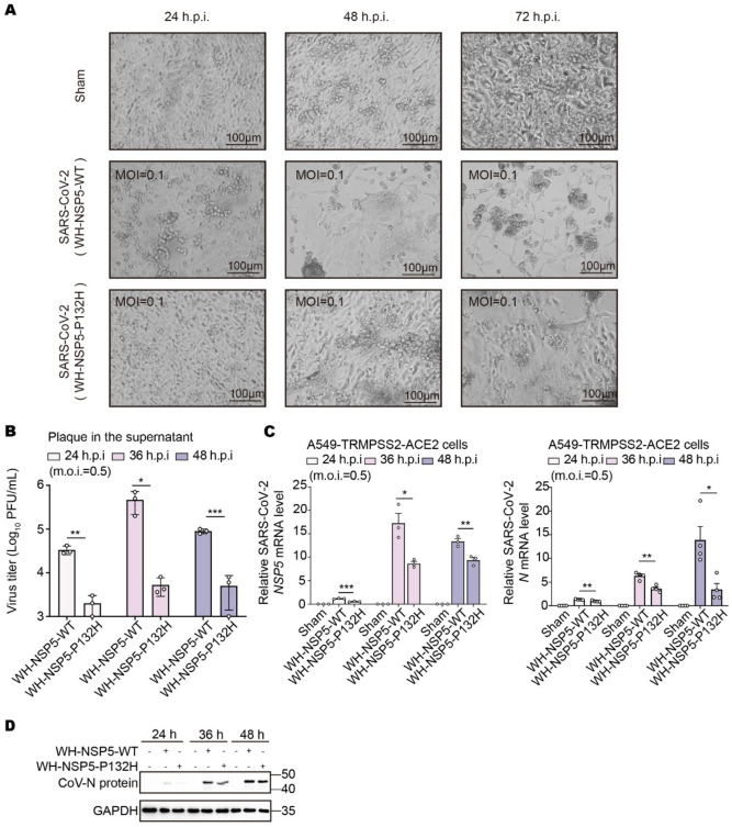
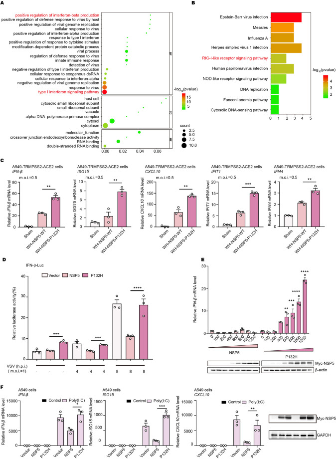
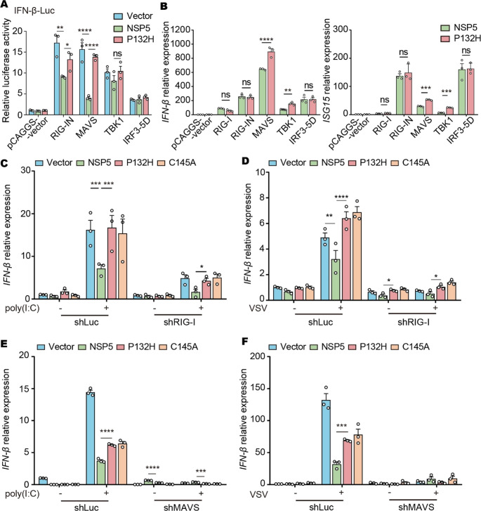
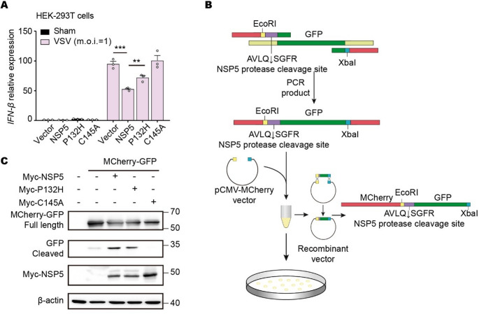
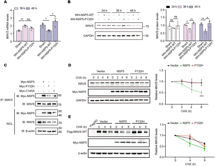
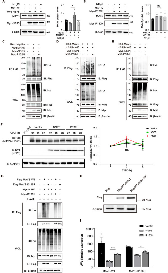
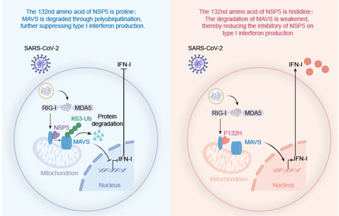
The prevalence of the Omicron variant of severe acute respiratory syndrome coronavirus 2 (SARS-CoV-2) is an important transition in the epidemic of coronavirus disease 2019 (COVID-19). Compared with other SARS-CoV-2 variants, Omicron and its subvariants exhibit decreased pathogenicity, thus contributing to the moderation of the epidemic. However, the mechanism underlying such changes is not fully understood. NSP5 is a SARS-CoV-2-encoded protease that counteracts antiviral immunity, and the P132H mutation of NSP5 is present exclusively in Omicron and its subvariants. In this study, we found that this mutation solely relieved cytopathogenicity and reduced the viral replication during SARS-CoV-2 infection. Further studies suggested that P132H blocked the NSP5-mediated degradation of MAVS by impairing the K136-linked ubiquitination of MAVS, thus restoring the IFN-β activation inhibited by NSP5. Structural analysis in silico suggested that P132H disrupted multiple hydrogen bonds between NSP5 and UbcH5b, an E2 ubiquitin-conjugating enzyme required for K136 ubiquitination. In summary, our results provide a potential mechanism explaining the decreased pathogenicity of the Omicron variant of SARS-CoV-2.

Keywords: IFN-β; MAVS; NSP5; P132H; SARS-CoV-2 variants.


Conflict of interest statement

Declarations. Competing interests: The authors have no relevant financial or non-financial interests to disclose.

Figures

Fig. 1
Fig. 1
The P132H mutation reduces the pathogenicity of NSP5. A Bright-field images of infected A549-TMPRSS2-ACE2 cells (m.o.i.= 0.1). Infected with the SARS-CoV-2 Wuhan strain NSP5 wild-type or the SARS-CoV-2 Wuhan strain P132H mutant virus, photos were taken 24, 48, and 72 h after infection. Sham, uninfected group. B Supernatants were collected after A549-TMPRSS2-ACE2 cells were infected with SARS-CoV-2 (m.o.i.=0.5) for 24, 36, and 48 h, and the virus titer in the supernatants was measured. C A549-TMPRSS2-ACE2 cells were mock infected or infected with the SARS-CoV-2 (m.o.i.=0.5) for 24, 36, and 48 h. The expression of the indicated the SARS-CoV-2 NSP5 or N protein genes was analyzed using qRT-PCR with total RNA as the template. D Immunoblotting analysis of A549-TMPRSS2-ACE2 cells infected with SARS-CoV-2 (m.o.i.= 0.5) for 24, 36, or 48 h. SARS-CoV-2 N protein antibody was used to analyze N protein expression. The data are presented as the means ± S.E.M.s. Significance was calculated using two-tailed, unpaired t-tests in Graphpad Prism 8.0. ****, P < 0.0001
Fig. 2
Fig. 2
P132H reduced the inhibition of type I interferon by NSP5. A, B NSP5 or P132H was transfected into HEK-293T cells and differentially expressed genes were screened through transcriptome sequencing. GO enrichment analysis (A). The GO database divides the functions of genes into three parts: cellular component (CC), molecular function (MF), and biological process (BP). Scatter plot of KEGG pathway enrichment statistics (B). The KEGG database, combines genomic information with highly organic combinations of hierarchical functional information and is used to analyze the signaling pathways of metabolic pathways, biological systems, and gene functions. The rich factor is the ratio of the number of differentially expressed genes annotated in this pathway term to the numbers of all genes annotated in this pathway term. A greater rich factor means greater intensiveness. A higher P value indicates greater intensiveness. C A549-TMPRSS2-ACE2 cells were either mock-infected or infected with SARS-CoV-2 (m.o.i.=0.5) for 36 h. The expression of the indicated IFN-β, ISG15, CXCL10, IFIT1, and IFI44 genes was analyzed via qRT-PCR via total RNA. D IFN-β luciferase reporters and Vector, NSP5 or P132H, were transfected into HEK-293T cells. The pRL-TK plasmid was transfected to normalize the transfection efficiency. The cells were further stimulated with VSV (m.o.i.=1), and samples were collected after 4 and 8 h of stimulation. E HEK-293 cells were transfected with the NSP5, or P132H plasmids of different concentrations as indicated 24 h later. The expression of the indicated IFN-β genes was analyzed via qRT-PCR via total RNA. The expression of NSP5 or P132H mutant in HEK-293T cells was detected by Western blotting via Myc tag antibody. F A549 cells were transfected with empty vector, NSP5, or P132H plasmids as indicated 24 h later. The cells were further stimulated with poly(I: C) 8 h. The cells were harvested to analyze the expression of IFN-β, ISG15, and CXCL10 using RT-qPCR. The expression of NSP5 or P132H mutant in HEK-293T cells was detected by Western blotting via Myc tag antibody. Significance was calculated via the two-tailed, unpaired t-tests in GraphPad Prism 8.0. *, P < 0.05; **, P < 0.01; ***, P < 0.001; ****, P < 0.0001. m.o.i. multiplicity of infection, h hours. The data are presented as the means ± SEMs
Fig. 3
Fig. 3
P132H targets MAVS to reduce the antagonistic effect of NSP5 on IFN-β. A Dual-luciferase reporter system to detect the effect of NSP5 or P132H on IFN-β promoter activation. Transfection of pRL-TK plasmid: IFN-β luciferase reporter plasmid = 1:10. Transfection of pRL-TK plasmid to normalize transfection efficiency. Co-transfection of vector (pEGFP-Myc), NSP5 or P132H plasmid and pCAGGS-vector, RIG-I-N, MAVS, TBK1 and IRF3-5D protein expression plasmids. Transfection was performed in HEK-293T cells. After 24 h of transfection, cells were lysed and luciferase activity was detected by dual luciferase assay. B HEK-293T cells were co-transfected with NSP5 or P132H and pCAGGS-vector, RIG-I, RIG-I N, MAVS, TBK1, and IRF3-5D protein expression plasmids. The expression of the indicated IFN-β and ISG15 genes was analyzed via qRT-PCR via total RNA. (C-D) qRT-PCR detection of IFN-β expression in RIG-I-knockdown cell lines expressing the Vector, NSP5, P132H or C145A plasmid. Transfection was performed for 24 h, followed by poly(I: C) stimulation (C). The cells were transfected for 30 h and stimulated with VSV for 8 h (D). (E-F) qRT-PCR detection of IFN-β expression in MAVS-knockdown cell lines expressing the Vector, NSP5, P132H or C145A plasmid. Transfection was performed for 24 h, followed by poly(I: C) stimulation (E). The cells were transfected for 30 h and stimulate with VSV for 8 h (F). The data are presented as the means ± S.E.M.s. Significance was calculated using two-tailed, unpaired t-tests in GraphPad Prism 8.0. *, P < 0.05; **, P < 0.01; ***, P < 0.001; ****, P < 0.0001
Fig. 4
Fig. 4
P132H does not affect on enzyme activity. A HEK-293T cells were transfected with empty vector, NSP5, P132H or C145A plasmids as indicated, 24 h later. The cells were further stimulated with VSV (m.o.i.=1). At the indicated time points, the cells were collected for RT-qPCR analysis to determine the relative expression levels of the target genes normalized to those of GAPDH. B A plasmid was constructed to validate the activity of the NSP5 protease. Using homologous recombination, an NSP5 protease cleavage sequence (AVLQSGFR) and a GFP fragment were inserted after the MCherry sequence of the vector pCMV-MCherry. C Immunoprecipitation detection of full-length GFP and cleaved fragments. The mCherry-GFP plasmid containing restriction sites was co-transfected with either the empty vector or the NSP5, P132H, or C145A plasmid into HEK-293T cells, and samples were collected after 36 h. The data are presented as the means ± S.E.M.s. Significance was calculated using two-tailed, unpaired t-tests in Graphpad Prism 8.0. **, P < 0.05; ***, P < 0.001
Fig. 5
Fig. 5
P132H reduces the degradation of MAVS by NSP5. A qRT-PCR analysis of A549-TMPRSS2-ACE2 cells infected with SARS-CoV-2 (m.o.i.= 0.5) for 36 and 48 h. B Immunoblotting analysis of A549-TMPRSS2-ACE2 cells infected with SARS-CoV-2 (m.o.i.= 0.5) for 24, 36, or 48 h. C NSP5, P132H and C145A interact with MAVS. HEK-293T cells were transfected with the indicated plasmids for 24 h before undergoing co-immunoprecipitation. The input and immunoprecipitates were immunoblotted with the indicated antibodies. The pEGFP-Myc vector was used to balance the total amount of DNA in each transfection. The immunoblotting results are representative of two independent experiments. D Immunoblotting analysis of WCLs from HEK-293T cells transfected with plasmids containing NSP5 or P132H and treated with DMSO or 100 µg/mL cycloheximide (CHX) and the indicated antibodies. MAVS was quantified by densitometry and plotted as shown on the right. E HEK-293T cells co-transfected with NSP5 or P132H and Flag-MAVS-WT were treated with DMSO or 100 µg/mL cycloheximide (CHX) for different durations and then analyzed by immunoblotting. The results represent one of three independent experiments. The data are presented as the means ± S.E.M.s. Significance was calculated using two-tailed, unpaired t-tests in Graphpad Prism 8.0. *, P < 0.05; **, P < 0.01
Fig. 6
Fig. 6
P132H weakens the inhibition of IFN by NSP5 by alleviating the polyubiquitination of MAVS.A, B Immunoblotting analysis of WCLs from HEK-293T cells transfected with plasmids containing NSP5 (A) and P132H (B) and treated with the proteasome inhibitor MG132 (25 µM) or the lysosome inhibitor NH4Cl (10 mM). C Immunoblotting analysis of the exogenous ubiquitination levels of MAVS and WCLs of in HEK-293T cells transfected with plasmids containing NSP5 or P132H and treated with the indicated antibodies. All the groups were treated with MG132 (20 µM) for 6 h before the samples were collected. D, E Immunoblotting analysis of the exogenous ubiquitination levels of MAVS and WCLs from HEK-293T cells transfected with plasmids containing NSP5 or P132H and ubiquitin (WT) and treated with K63(D) or K48(E) and the indicated antibodies. Ub, ubiquitin. All the groups were treated with MG132 (20 µM) for 6 h before the samples were collected. F Immunoblotting analysis of WCLs of HEK-293T cells stably expressing MAVS-K136R transfected with plasmids containing NSP5 or P132H, treated with DMSO or cycloheximide (CHX, 100 µg/ml), and treated with the indicated antibodies. MAVS protein was quantified by densitometry and plotted as shown on the right. G Immunoblotting analysis of the levels of ubiquitinated MAVS and WCLs in HEK-293T cells overexpressing MAVS-WT or MAVS-K136R transfected with plasmids containing NSP5 or P132H and treated with the indicated antibodies. All the groups were treated with MG132 (20 µM) for 6 h before the samples were collected. H Immunoblot analysis of WCLs transfected with MAVS WT or MAVS-K136R in MAVS-knockdown cell lines. I The mRNA levels of IFN-β in total RNA extracted from cells expressing MAVS-WT or MAVS-K136R in the presence or absence of NSP5 or P132H were analyzed by qRT-PCR. The data are presented as the means ± S.E.M.s. Significance was calculated via two-tailed, unpaired t-tests in GraphPad Prism 8.0. *, P < 0.05; ***, P < 0.001
Fig. 7
Fig. 7
NSP5, as an E3 ligase, promotes the polyubiquitination-mediated degradation of MAVS, blocks downstream signal transmission, inhibits IFN-β induction, and promotes viral immune escape. After the NSP5 protease mutation, specifically the P132H mutation, the degradation of MAVS was weakened and the induction of IFN was promoted

Similar articles

References

    1. Sigal A, Milo R, Jassat W (2022) Estimating disease severity of Omicron and Delta SARS-CoV-2 infections. Nat Rev Immunol 22:267–269. 10.1038/s41577-022-00720-5 - PMC - PubMed
    1. Diamond MS, Kanneganti T-D (2022) Innate immunity: the first line of defense against SARS-CoV-2. Nat Immunol 23:165–176. 10.1038/s41590-021-01091-0 - PMC - PubMed
    1. Rehwinkel J, Gack MU (2020) RIG-I-like receptors: their regulation and roles in RNA sensing. Nat Rev Immunol 20:537–551. 10.1038/s41577-020-0288-3 - PMC - PubMed
    1. He Q-q, Huang Y, Nie L et al (2023) MAVS integrates glucose metabolism and RIG-I-like receptor signaling. Nat Commun 14:5343. 10.1038/s41467-023-41028-9 - PMC - PubMed
    1. Minkoff JM, tenOever B (2023) Innate immune evasion strategies of SARS-CoV-2. Nat Rev Microbiol 21:178–194. 10.1038/s41579-022-00839-1 - PMC - PubMed

Substances

Supplementary concepts

LinkOut - more resources