. 2025 Jul 30;82(1):292.
doi: 10.1007/s00018-025-05821-7.

Proteome of oocyte spindle identifies Ccdc69 regulates spindle assembly like "band-tightening spell"

Affiliations

Proteome of oocyte spindle identifies Ccdc69 regulates spindle assembly like "band-tightening spell"

Jia-Ni Guo et al. Cell Mol Life Sci. .

Abstract

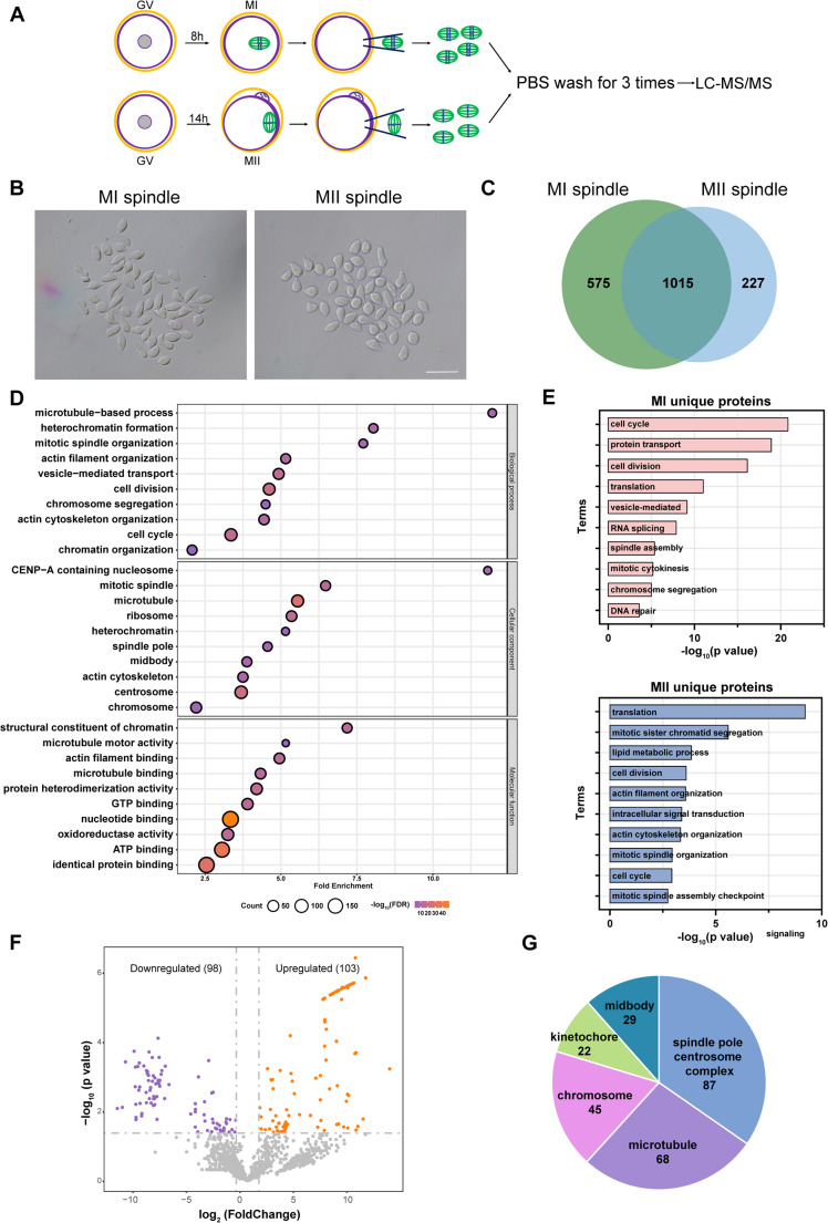
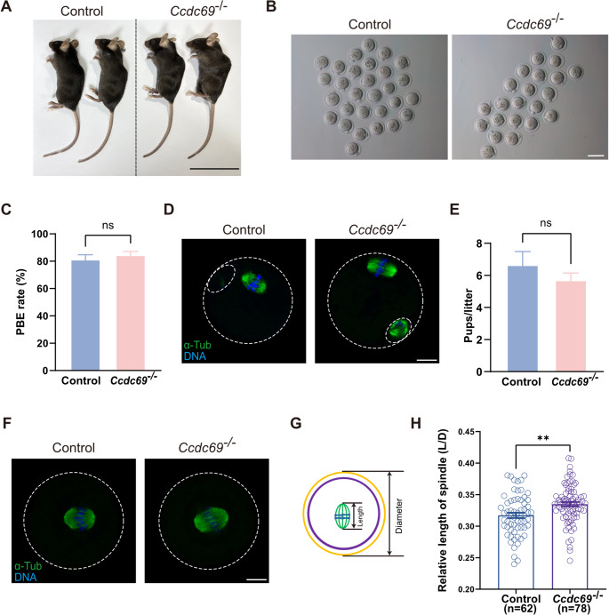
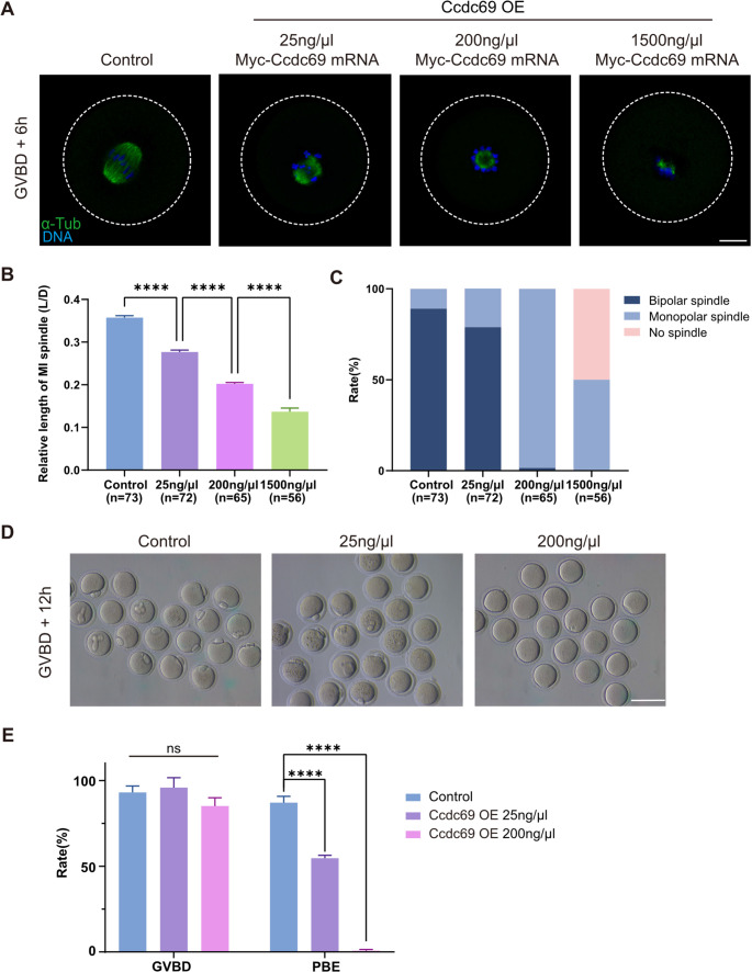
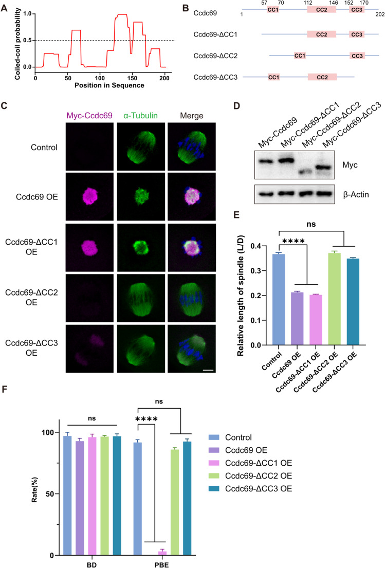
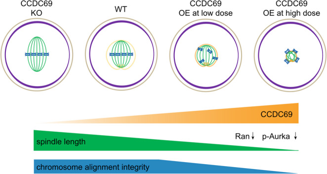
Meiotic spindle is an intricate structure and required for chromosome segregation and the proper meiotic progression during oocyte maturation, and its function is regulated by a complex network of proteins located at spindle and its peripheral region. However, proteome of meiotic spindle remains poorly characterized. Here, we acquired the proteomic profile of spindles isolated from metaphase I (MI) and metaphase II (MII) mouse oocytes. In particular, we identified Ccdc69 as a novel regulator of spindle assembly in mouse oocytes. Although deletion of Ccdc69 did not affect female fertility, the MI spindles were elongated in Ccdc69 knockout oocytes. Overexpression of Ccdc69 induced spindle defects by reducing microtubule formation and disturbing acentriolar microtubule organization centers (aMTOCs) distribution. Furthermore, Ccdc69 overexpression impaired kinetochore-microtubule (K-MT) attachment and delayed meiotic progression by abnormal activation of spindle assembly checkpoint (SAC). Taken together, our study depicts the proteome of spindles during mouse oocyte maturation and demonstrates that Ccdc69 regulates spindle assembly and meiotic progression the way similar to "The Tightening Spell of Sun Wukong's Golden Headband" in the famous Chinese Classic Journey to the West.

Keywords: Ccdc69; Meiosis; Oocyte; Proteome; Spindle assembly.


Conflict of interest statement

Declarations. Ethics approval and consent to participate: Mice were feed in specific pathogen-free barrier facilities at experimental animal center of the Institute of Zoology (IOZ), Chinese Academy of Science (Beijing, China). All animal handling protocols were approved by the guidelines of Animal Research Committee of the Institute of Zoology. Consent for publication: Not applicable. Conflict of interest: The authors declare that they have no conflict of interest.

Figures

Fig. 1
Fig. 1
Proteomic profiling of the meiotic spindles of mouse oocytes. A. Schematic overview of the workflow for proteomic profiling of meiotic spindles isolated from mouse oocytes at MI and MII stage respectively. B. Representative images of meiotic spindles from mouse oocytes at MI and MII stage respectively. Scale bar = 50 μm. C. Venn diagram showing the numbers of proteins identified at different stages. D. Gene ontology (GO) enrichment analysis of shared proteins of spindles from mouse oocytes at MI and MII stages. E. GO enrichment analysis of specific proteins of MI and MII spindles respectively. F. Volcano plot showing DEPs (downregulated proteins, purple; upregulated proteins, orange) in the shared proteins of MI and MII spindles G. Pie chart depicting the distribution of proteins among the spindles
Fig. 2
Fig. 2
Deletion of Ccdc69 elongates MI spindles but does not affect female fertility. A. Macroscopic phenotype of the control and Ccdc69−/− female mice. Scale bar = 5 cm. B. Representative images of MII oocytes collected from control and Ccdc69−/− female mice. Scale bar = 100 μm. C. Comparison of PBE rates of control oocytes and Ccdc69−/− oocytes in vivo. p = 0.5165, n = number of oocytes. D. Normal spindle assembly and chromosome alignment in control and Ccdc69−/− MII oocytes. Green, α-Tubulin; blue, DNA. The small circle represented polar body. Scale bar = 20 μm. E. Female Ccdc69−/− mouse were fertile. At least three mice of each genotype were examined. p = 0.3845. F. Elongated spindles in control and Ccdc69−/− MI oocytes. Green, α-Tubulin; blue, DNA. Scale bar = 20 μm. G. Scheme of measuring the relative length of spindle. H. Relative length of spindles in MI oocytes from control and Ccdc69−/− female mouse. **p < 0.01, n = number of oocytes
Fig. 3
Fig. 3
Overexpression of Ccdc69 shortens spindles and impairs meiotic progression. A. Representative images of spindle morphology at MI stage in control and Ccdc69-OE oocytes microinjected with different concentrations of Myc-Ccdc69 mRNAs. Green, α-Tubulin; blue, DNA. Scale bar = 20 μm. B. Relative length of spindles at MI stage in control and Ccdc69-OE oocytes microinjected with different concentrations of Myc-Ccdc69 mRNAs. ****p < 0.0001, n = number of oocytes. C. Percentages of different types of spindles at MI stage in the control and Ccdc69-OE oocytes with different concentrations of Myc-Ccdc69 mRNAs. n = number of oocytes. D. Representative images of the occurrence of PBE in control and Ccdc69-OE oocytes microinjected with different concentrations of Myc-Ccdc69 mRNAs. Scale bar = 100 μm. E. Percentages of control and Ccdc69-OE oocytes that underwent GVBD and PBE. ****p < 0.0001
Fig. 4
Fig. 4
Ccdc69 regulates microtubule generation by mediating Ran. A. Representative images of Myc-Ccdc69 localization during microtubule formation. Magenta, Myc-Ccdc69; green, α-Tubulin; blue, DNA. Scale bar = 10 μm. White arrows indicate that microtubules are surrounded by Ccdc69. GV, GVBD, GV + 4 h: oocytes microinjected with 200ng/µl Myc-Ccdc69 mRNAs. GV + 8 h: oocytes microinjected with 25ng/µl Myc-Ccdc69 mRNAs. B. Representative images of spindles at different times after released from IBMX in control and Ccdc69-OE oocytes (200ng/µl Myc-Ccdc69 mRNAs). Green, α-Tubulin; blue, DNA. Scale bar = 10 μm. C. Relative intensity of α-Tubulin in control and Ccdc69-OE oocytes at different times after released from IBMX. At least 30 oocytes of control or overexpression oocytes were examined at each time. ****p < 0.0001. D. Expression level of Ran in control and Ccdc69-OE oocytes at MI stage (100 oocytes per lane) was determined by western blotting. Quantity analysis was performed with three independent experiments. ***p < 0.001
Fig. 5
Fig. 5
Overdosed Ccdc69 impairs aMTOCs distribution and spindle bipolarity. A. Relative length of spindles in control and Ccdc69-OE oocytes at different times after released from IBMX. At least 35 oocytes of control or overexpression group were examined at each timepoint. ***p < 0.001, ****p < 0.0001. B. Representative images of aMTOCs distribution during spindle formation in control and Ccdc69-OE oocytes. Red, γ-Tubulin; blue, DNA. Scale bar = 10 μm. C. Representative images of p-Aurka in control and Ccdc69-OE oocytes at MI stage. Red, p-Aurka; green, α-Tubulin; blue, DNA. Scale bar = 10 μm. D. Immunofluorescence analysis of p-Aurka in control and Ccdc69-OE oocytes. ****p < 0.0001, n = number of oocytes. E. Representative images of p-Aurka in control and Ccdc69−/− oocytes at MI stage. Red, p-Aurka; green, α-Tubulin; blue, DNA. Scale bar = 10 μm. F. Immunofluorescence analysis of p-Aurka in control and Ccdc69-OE oocytes. **p < 0.01, n = number of oocytes. G. Representative images of Tpx2 in control and Ccdc69-OE oocytes at MI stage. Red, Tpx2; green, α-Tubulin; blue, DNA. Scale bar = 10 μm. H. Representative images of Tacc3 in control and Ccdc69-OE oocytes at MI stage. Magenta, Tacc3; green, α-Tubulin; blue, DNA. Scale bar = 10 μm
Fig. 6
Fig. 6
Excess Ccdc69 disrupts chromosome alignment and SAC function. A. Statistical analysis of PB1 extrusion time in control and Ccdc69-OE (25 ng/µl Myc-Ccdc69 mRNAs) oocytes. ***p < 0.001, n = number of oocytes. B. Representative images of chromosome alignment in control and Ccdc69-OE oocytes at MI stage. Green, α-Tubulin; blue, DNA. Scale bar = 10 μm. C. Rates of chromosome misalignment in control and Ccdc69-OE oocytes at MI stage. *p < 0.05, n = number of oocytes. D. Representative images of K–MT attachments, including amphitelic attachments, merotelic attachments, and lateral interactions, in control and Ccdc69-OE oocytes at MI stage. Red, ACA; blue, DNA. Scale bar = 10 μm. E. Rates of erroneous K–MT attachments in control and Ccdc69-OE oocytes at MI stage. **p < 0.01, n = number of oocytes. F. Representative images of SAC activity as indicated by localization of Bub3 at late MI (7 h after GVBD) in control and Ccdc69-OE oocytes. Red, ACA; green, Bub3; blue, DNA. Scale bar = 10 μm. G. Immunofluorescence analysis of bub3 in control and Ccdc69-OE oocytes. The relative intensity of Bub3 was compared to the intensity of ACA. ****p < 0.0001, n = number of kinetochores. H. Representative time-lapse images showing the degradation of Securin-mCherry in control and Ccdc69-OE oocytes after GVBD. Images are overlaid with mCherry and GFP channels. Red, Securin; green, H2B. Scale bar = 20 μm. I. Fluorescence intensity of Securin-mCherry in control and Ccdc69-OE oocytes captured every 30 min. ****p < 0.0001, n = number of oocytes
Fig. 7
Fig. 7
Roles of the Coiled-Coil domains in Ccdc69. A. The PCOILS program predicts that mouse Ccdc69 contains three coiled-coil domains (amino acids 57–70, 112–146 and 152–170). B. Schematic diagram of truncated Ccdc69. C. Representative images of truncated Myc-Ccdc69 in oocytes at MI stage. Magenta, Myc-Ccdc69; green, α-Tubulin; blue, DNA. Scale bar = 10 μm. D. Expression level of truncated Myc-Ccdc69 was determined by western blotting. The plasmids of each truncated Myc-Ccdc69 were transfected into HEK293T cells to culture for 48 h. E. Relative length of spindles in MI oocytes from control (n = 58), Ccdc69-OE (n = 42), Ccdc69-ΔCC1-OE (n = 50), Ccdc69-ΔCC2 (n = 35) and Ccdc69-ΔCC3 (n = 53) groups. ****p < 0.0001, n = number of oocytes. F. Percentage of control, Ccdc69-OE, Ccdc69-ΔCC1-OE, Ccdc69-ΔCC2-OE and Ccdc69-ΔCC3-OE oocytes that underwent GVBD and PBE. ****p < 0.0001. The concentration of each mRNA was 200 ng/µl
Fig. 8
Fig. 8
Schematic diagram of the roles of Ccdc69 in mouse oocytes. Ccdc69 localizes around the spindle and works as a band-tightening spell to prevent excessive spindle extension. Ccdc69 lessens spindles by reducing Ran and disrupts bipolarity by regulating p-Aurka. The spindle length and chromosome alignment integrity decrease with the increase of Ccdc69

Similar articles

References

    1. Thomas C, Cavazza T, Schuh M (2021) Aneuploidy in human eggs: contributions of the meiotic spindle. Biochem Soc Trans 49:107–118 - PMC - PubMed
    1. Gruhn JR, Zielinska AP, Shukla V, Blanshard R, Capalbo A, Cimadomo D et al (2019) Chromosome errors in human eggs shape natural fertility over reproductive life span. Science 365:1466–1469 - PMC - PubMed
    1. Dumont J, Desai A (2012) Acentrosomal spindle assembly and chromosome segregation during oocyte meiosis. Trends Cell Biol 22:241–249 - PMC - PubMed
    1. Szollosi D, Calarco P, Donahue RP (1972) Absence of centrioles in the first and second meiotic spindles of mouse oocytes. J Cell Sci 11:521–541 - PubMed
    1. Manandhar G, Schatten H, Sutovsky P (2005) Centrosome reduction during gametogenesis and its significance. Biol Reprod 72:2–13 - PubMed

LinkOut - more resources