Multicenter Study
. 2025 Sep 1;160(9):973-982.
doi: 10.1001/jamasurg.2025.2493.

Exosomal Liquid Biopsy for the Early Detection of Gastric Cancer: The DESTINEX Multicenter Study

Affiliations
Multicenter Study

Exosomal Liquid Biopsy for the Early Detection of Gastric Cancer: The DESTINEX Multicenter Study

Silei Sui et al. JAMA Surg. .

Abstract

Importance: Gastric cancer (GC) is the third leading cause of cancer-related deaths worldwide, primarily attributed to delayed detection. The invasive and cost-prohibitive nature of endoscopy for GC screening highlights the urgent need for noninvasive biomarkers.

Objective: To develop an exosome-based diagnostic signature to facilitate blood-based, early detection of patients with GC.

Design, setting, and participants: This was a multicenter, population-based, retrospective, case-control study that analyzed specimens collected between January 1, 2016, and December 30, 2020. The study encompassed the discovery, training, validation, and evaluation phases of biomarker development. This study was conducted at 4 major referral centers: Nagoya University Hospital in Japan and Ajou University Hospital, Asan Medical Center, and Samsung Medical Center in South Korea, providing a broad representation of advanced clinical care settings. The study included patients with GC, classified according to the TNM (tumor-node-metastasis) classification (8th edition), and controls without disease. Key sociodemographic data, including age and sex, were recorded for all participants. Data were analyzed from October 2022 to July 2024.

Exposures: Results were obtained from tissue and serum microRNA (miRNA) profiling and expression analysis. Frozen tissue collection and blood draws were conducted intraoperatively, preoperatively, and 3 months postoperatively.

Main outcomes and measures: Diagnostic performance of GC detection using an exosome-based miRNA signature.

Results: A total of 809 specimens from 480 patients (mean [SD] age, 61.9 [9.8] years; 336 male [70%]) in the training and validation cohorts were analyzed. A panel of 8 cell-free miRNAs and 10 exosomal miRNAs was initially developed in the discovery phase, which was subsequently reduced using machine learning algorithms to a panel of 8 cell-free and 9 exosomal miRNAs during the training phase. This 17-miRNA signature robustly identified GC with area under the curve (AUC) values of 96.3% (95% CI, 94.3%-98.4%) and 95.3% (95% CI, 92.8%-97.9%) in the training and validation cohorts, respectively. Additionally, 5 overlapping miRNAs were observed between cell-free and exosomal panels and exhibited a comparable efficacy in identifying patients with GC. Finally, we established a 10-miRNA signature (Destinex), which successfully identified early-stage (pT1) GC (AUC = 96.8%; 95% CI, 93.5%-100%). Finally, a significant decrease in miRNA expression levels in postsurgery serum specimens confirmed the robustness of the panel specificity.

Conclusion and relevance: Results of this case-control study suggest that the Destinex assay was robust for early detection of GC, highlighting its potential for clinical application in the noninvasive identification of GC.


Conflict of interest statement

Conflict of Interest Disclosures: Dr Goel reported having a patent pending outside the submitted work. No other disclosures were reported.

Figures

Figure 1.
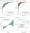
Figure 1.. Genome-Wide Discovery of MicroRNA (miRNA) Candidates for Detection of Gastric Cancer
A, Venn diagram shows the overlapping differentially expressed miRNAs among tissue, cell-free (cf), and exosomes (exo). The Vennerable package, version 3.1, in R software (R Project for Statistical Computing) was used to create the Venn diagram. B, The heatmap depicts the 10 overlapping upregulated miRNAs between tissue and exosomes (exo-miRNAs). C, The heatmap illustrates the 8 overlapping upregulated miRNAs between tissue and cell-free (cf-miRNAs). D, Receiver operating characteristic curve analysis evaluates the performance of 8 cf-miRNA panels and 10 exo-miRNA panels that robustly distinguished gastric cancer from controls without gastric cancer. ROC curves are shown as 95% CIs. AUC indicates area under the curve; FC, fold change; hsa, Homo sapiens (human miRNA).
Figure 2.
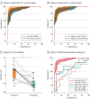
Figure 2.. Development of a Noninvasive MicroRNA (miRNA)–Based Signature for Detection of Patients With Gastric Cancer (GC)
A, Receiver operating characteristic (ROC) curve analysis evaluates the performance of 8 cell-free miRNA (cf-miRNA) panel and 9 exosomal miRNA (exo-miRNA) panel in the training cohort. ROC curves are shown as 95% CIs. B, ROC curve analysis illustrates the performance of the 17-miRNA combination signature in the training and validation cohort. ROC curves are shown as 95% CIs. C, Odds ratios for GC with restricted cubic splines as a function of risk score values (95% CIs for odds ratios are presented as dashed lines). D, The decision curve shows the net benefit for the cf-miRNA, exo-miRNA, and combination signature in patients with GC from the validation cohort. AUC indicates area under the curve.
Figure 3.
Figure 3.. Construction of a Clinically Feasible Signature for Noninvasive Detection of Patients With Gastric Cancer (GC)
A, Receiver operating characteristic (ROC) curve analysis illustrates the performance of the Destinex assay in the training and validation cohort. B, The sensitivity and specificity of 5 cell-free microRNA (cf-miRNA) panel, 5 exosomal miRNA (exo-miRNA) panel, and the Destinex assay were shown in the validation cohort. C, The decision curve analysis reveals the net benefit of the Destinex assay and 17-miRNA combination signatures in patients with GC from the validation cohort. The Destinex assay indicates detection of GC by combining 5 cf-miRNAs and exo-miRNAs. D, The comparison of accuracy, precision, recall, and F1 score between the Destinex assay and the 17-miRNA combination signature is shown in the validation cohort. AUC indicates area under the curve. aP < .05. bP < .001.
Figure 4.
Figure 4.. The Noninvasive Destinex Assay Efficiently Identifies Patients With Early Gastric Cancer (GC)
A, Receiver operating characteristic (ROC) curve analysis reveals the performance of the Destinex assay in patients with the pathological tumor stage 1 (pT1) and pT2-4 stages from the validation cohort. B, ROC curve analysis reveals the performance of the Destinex assay in patients with stages I-II and III-IV from the validation cohort. ROC curves are shown as 95% CIs. C, Assessment of risk probability based on the Destinex assay between presurgery and postsurgery GC samples. D, ROC curve analysis shows the performance of the Destinex assay in different gastrointestinal malignancies (GC, colorectal cancer [CRC], pancreatic ductal adenocarcinoma cancer [PDAC], esophageal squamous cell carcinoma [ESCC], intrahepatic cholangiocarcinoma [ICC], hepatocellular carcinoma [HCC]). The Destinex assay indicates detection of GC by combining 5 cell-free microRNAs (cf-miRNAs) and exosomal miRNAs (exo-miRNAs). AUC indicates area under the curve. aP < .001.

Comment on

References

    1. Smyth EC, Nilsson M, Grabsch HI, van Grieken NC, Lordick F. Gastric cancer. Lancet. 2020;396(10251):635-648. doi: 10.1016/S0140-6736(20)31288-5 - DOI - PubMed
    1. Sung H, Ferlay J, Siegel RL, et al. Global Cancer Statistics 2020: GLOBOCAN estimates of incidence and mortality worldwide for 36 cancers in 185 countries. CA Cancer J Clin. 2021;71(3):209-249. doi: 10.3322/caac.21660 - DOI - PubMed
    1. Lordick F, Carneiro F, Cascinu S, et al. ; ESMO Guidelines Committee. Electronic address: clinicalguidelines@esmo.org . Gastric cancer: ESMO clinical practice guideline for diagnosis, treatment and follow-up. Ann Oncol. 2022;33(10):1005-1020. doi: 10.1016/j.annonc.2022.07.004 - DOI - PubMed
    1. Pasechnikov V, Chukov S, Fedorov E, Kikuste I, Leja M. Gastric cancer: prevention, screening and early diagnosis. World J Gastroenterol. 2014;20(38):13842-13862. doi: 10.3748/wjg.v20.i38.13842 - DOI - PMC - PubMed
    1. Lee JH, Kim JG, Jung HK, et al. Clinical practice guidelines for gastric cancer in Korea: an evidence-based approach. J Gastric Cancer. 2014;14(2):87-104. doi: 10.5230/jgc.2014.14.2.87 - DOI - PMC - PubMed