

Differentiation of HL-60 cells into primed neutrophils for the evaluation of antiapoptotic effects of poorly soluble nanoparticles
- PMID: 40737277
- PMCID: PMC12309983
- DOI: 10.1371/journal.pone.0328717
Differentiation of HL-60 cells into primed neutrophils for the evaluation of antiapoptotic effects of poorly soluble nanoparticles
Abstract
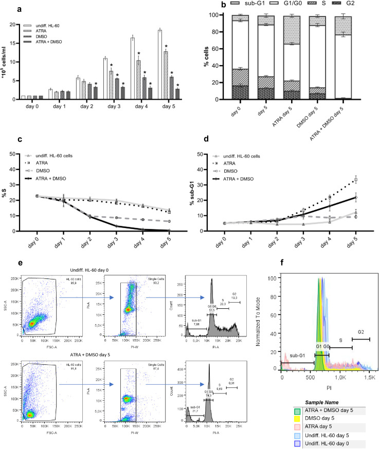
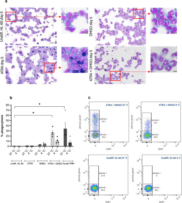
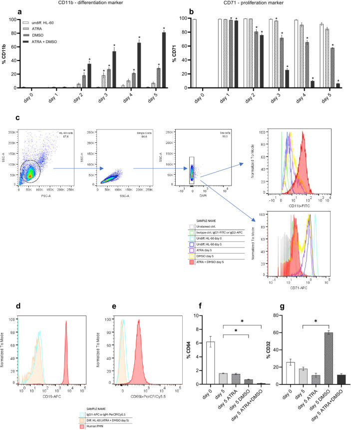
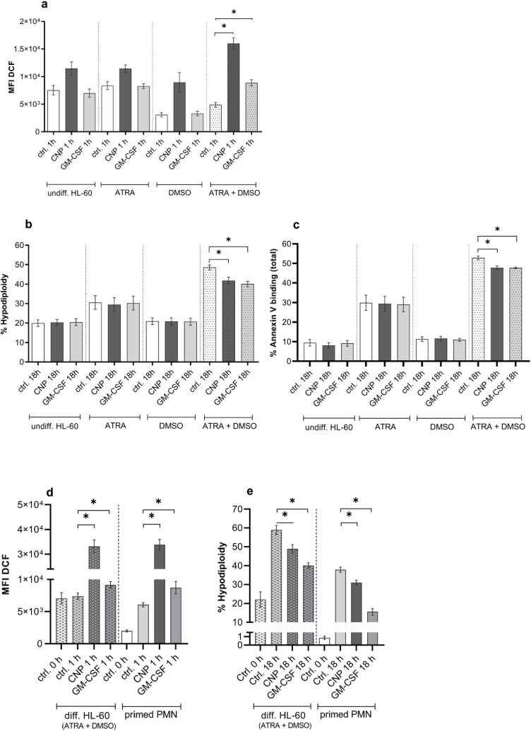
Background: Neutrophil apoptosis is an important determinant of intensity and duration of neutrophilic inflammation. The interaction of poorly soluble nanoparticles with primed neutrophils can reduce their natural apoptosis rates. This reaction may contribute to pathogenic consequences of increased neutrophilic inflammation. Toxicological studies aiming to identify hazards of such materials with primary neutrophils are however challenging due to the short life span of these cells and a high donor to donor variability. Our purpose was the establishment of a culturable neutrophil-like cell line as a suitable model for studies of antiapoptotic effects of poorly soluble combustion-derived environmental nanoparticles. Therefore, differentiation protocols for the myeloid HL-60 cell line based on commonly used differentiation inducers all-trans retinoic acid (ATRA) and dimethyl sulfoxide (DMSO) were established and compared.
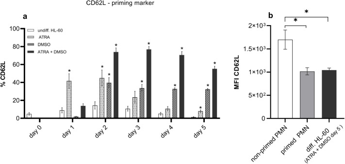
Results: The data demonstrate that only a combined cell treatment with ATRA and DMSO for a period of 5 days leads to the complete HL-60 differentiation with the typical neutrophil morphology and characteristic features of neutrophil maturation like cell cycle arrest, increase in differentiation marker CD11b, loss of proliferation marker CD71, and increased phagocytic capacity. Exposure of cells differentiated with ATRA + DMSO to carbon nanoparticles or proinflammatory cytokine granulocyte-macrophage colony-stimulating factor (GM-CSF) revealed a delay of apoptosis causally linked to intracellular reactive oxygen species (ROS). These data verified our earlier findings with human peripheral primed neutrophils from donors with slightly elevated proinflammatory blood plasma factors. Moreover, completely differentiated HL-60 cells possessed similar levels of L-selectin CD62L as neutrophils with primed immunophenotype, thus representing the biology of primed inflammatory neutrophils.
Conclusion: Neutrophil-like HL-60 cells differentiated according to our protocol could be an appropriate substitute cell line model for studies on the effects of inhalable nanomaterials on primed inflammatory neutrophils like lung neutrophils. For such toxicological studies our cell model is preferable to peripheral neutrophils, as blood neutrophils not always occur in a primed state and primed lung neutrophils from donors are not available for this purpose.
Copyright: © 2025 Hornstein, Unfried. This is an open access article distributed under the terms of the Creative Commons Attribution License, which permits unrestricted use, distribution, and reproduction in any medium, provided the original author and source are credited.
Conflict of interest statement
The authors have declared that no competing interests exist.
Figures
Similar articles
-
Sexual Harassment and Prevention Training.2024 Mar 29. In: StatPearls [Internet]. Treasure Island (FL): StatPearls Publishing; 2025 Jan–. 2024 Mar 29. In: StatPearls [Internet]. Treasure Island (FL): StatPearls Publishing; 2025 Jan–. PMID: 36508513 Free Books & Documents.
-
Positron emission tomography-adapted therapy for first-line treatment in individuals with Hodgkin lymphoma.Cochrane Database Syst Rev. 2015 Jan 9;1(1):CD010533. doi: 10.1002/14651858.CD010533.pub2. Cochrane Database Syst Rev. 2015. Update in: Cochrane Database Syst Rev. 2025 Mar 26;3:CD010533. doi: 10.1002/14651858.CD010533.pub3. PMID: 25572491 Free PMC article. Updated.
-
G-CSF and GM-CSF for treating or preventing neonatal infections.Cochrane Database Syst Rev. 2003;2003(3):CD003066. doi: 10.1002/14651858.CD003066. Cochrane Database Syst Rev. 2003. PMID: 12917944 Free PMC article.
-
Management of urinary stones by experts in stone disease (ESD 2025).Arch Ital Urol Androl. 2025 Jun 30;97(2):14085. doi: 10.4081/aiua.2025.14085. Epub 2025 Jun 30. Arch Ital Urol Androl. 2025. PMID: 40583613 Review.
-
Ambulatory Oxygen for Pulmonary Fibrosis (OxyPuF): a randomised controlled trial and acceptability study.Health Technol Assess. 2025 Jul 2:1-33. doi: 10.3310/TWKS4194. Online ahead of print. Health Technol Assess. 2025. PMID: 40613728
References
MeSH terms
Substances
LinkOut - more resources
Full Text Sources
Research Materials
