. 2025 Jul 30;20(7):e0325767.
doi: 10.1371/journal.pone.0325767. eCollection 2025.

Role of rosuvastatin and pitavastatin in alleviating diabetic cardiomyopathy in rats: Targeting of RISK, NF-κB/ NLRP3 inflammasome and TLR4/ NF-κB signaling cascades

Affiliations

Role of rosuvastatin and pitavastatin in alleviating diabetic cardiomyopathy in rats: Targeting of RISK, NF-κB/ NLRP3 inflammasome and TLR4/ NF-κB signaling cascades

Dalia O Saleh et al. PLoS One. .

Abstract

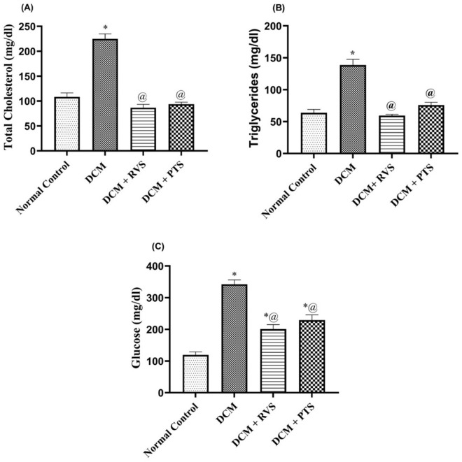
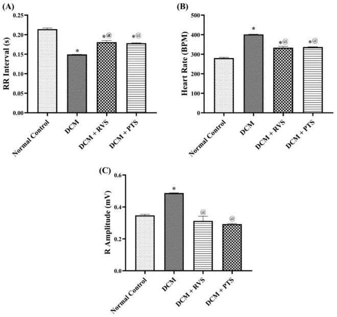
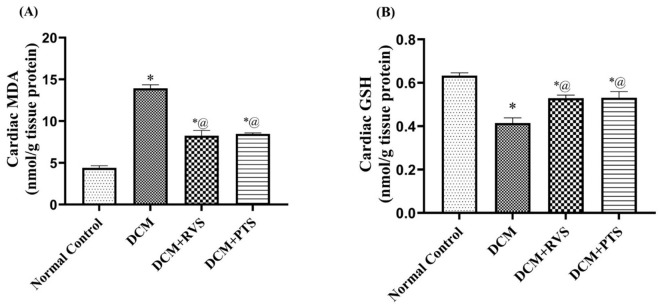
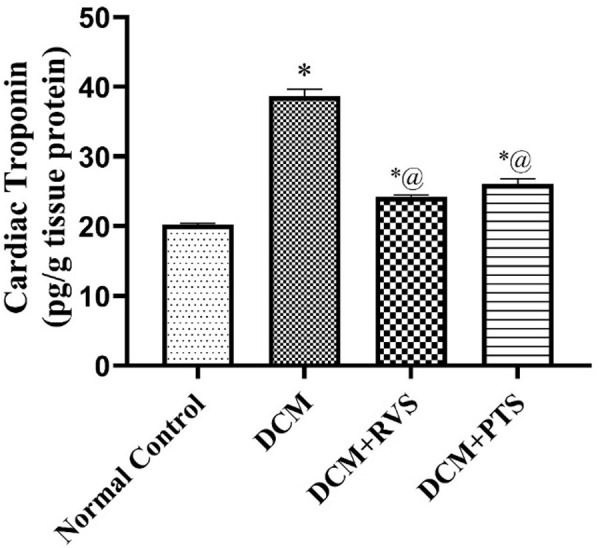
Diabetic cardiomyopathy (DCM) is a serious outcome of type II diabetes mellitus (T2DM) and a key contributor to high morbidity and death in diabetic individuals. The current research is intended to elucidate and compare the therapeutic benefits of rosuvastatin (RVS) and pitavastatin (PTS) in mitigating DMC-induced in rats and exploring the possible underlying molecular signaling pathways. DCM was prompted by feeding rats a high-fat/fructose (F/Fr) diet for eight weeks with a sub-diabetogenic dose of streptozotocin (35 mg/kg; i.p) injection at week seven. All rats were allocated into four groups: a normal control group, a DCM-induced positive control group, the RVS group of DCM-induced rats that were treated once daily with 10 mg/kg of RVS, and the PTS group of DCM rats that were treated with 0.8 mg/kg of PTS. Rats were given the treatments orally for four consecutive weeks. The outcome of the existing work discovered that RVS and PTS significantly improved T2DM-associated DCM, as evidenced by the amelioration of glucose, lipids, cardiac markers, ECG parameters, and redox status. Considering the relationship between oxidative stress and inflammation, this attenuation was evidenced by the downregulation of redox, inflammatory, and cellular fibrotic cascades, namely RISK, NF-κB/NLRP3 inflammasome, and TLR4/NF-κB signaling pathways. Additionally, the histopathological examinations confirmed these structural alterations in the myocardium. Besides, RVS and PTS diminished the expression of caspase-1 assessed by immunochemical staining. In summary, the present study demonstrated that RVS and PTS mitigated the metabolic abnormalities associated with T2DM-induced DCM.


Conflict of interest statement

The authors have declared that no competing interests exist.

Figures

Fig 1
Fig 1. Effect of Rosuvastatin and Pitavastatin on metabolic biomarkers; Cholesterol(A), Total Triglycerides (B), and Glucose (C) against DCM-induced in Rats.
Each bar represents the mean ± SEM of 6 rats. * Vs normal control group & @ vs DCM control group at p < 0.05. DCM, diabetic cardiomyopathy; RVS, Rosuvastatin; PTS, Pitavastatin.
Fig 2
Fig 2. Effect of Rosuvastatin and Pitavastatin on Cardiac Contractility measurements; RR Interval (A), Heart Rate (B), and R Amplitude (C) against DCM-induced in Rats.
Each bar represents the mean ± SEM of 6 rats. * Vs normal control group & @ vs DCM control group at p < 0.05. DCM, diabetic cardiomyopathy; RVS, Rosuvastatin; PTS, Pitavastatin.
Fig 3
Fig 3. Effect of Rosuvastatin and Pitavastatin on Cardiac Conductivity and Rhythmicity measurements PR Interval (A), QRS Interval (B), QTc (C), and ST Height (D) against DCM-induced Rats.
Each bar represents the mean ± SEM of 6 rats. * Vs normal control group & @ vs DCM control groupat p < 0.05. DCM, diabetic cardiomyopathy; RVS, Rosuvastatin; PTS, Pitavastatin.
Fig 4
Fig 4. Effect of Rosuvastatin and Pitavastatin on oxidative stress-related biomarkers; MDA (A) and GSH (B) against diabetic cardiomyopathy-induced in Rats.
Each bar represents the mean ± SEM of 6 rats. * Vs normal control group & @ vs DCM control group at p < 0.05. DCM, diabetic cardiomyopathy; RVS, Rosuvastatin; PTS, Pitavastatin.
Fig 5
Fig 5. Effect of Rosuvastatin and Pitavastatin on cardiac NLRP3 inflammasome (A) and pro-fibrotic IL-1 β (B) against diabetic cardiomyopathy-induced in Rats.
Each bar represents the mean ± SEM of 6 rats. * Vs normal control group & @ vs DCM control group at p < 0.05. DCM, diabetic cardiomyopathy; RVS, Rosuvastatin; PTS, Pitavastatin.
Fig 6
Fig 6. Effect of Rosuvastatin and Pitavastatin on cardiac Akt (A) and p-GSK-3β (B) against diabetic cardiomyopathy-induced in Rats.
Each bar represents the mean ± SEM of 6 rats. * Vs normal control group & @ vs DCM control group at p < 0.05. DCM, diabetic cardiomyopathy; RVS, rosuvastatin; PTS, pitavastatin.
Fig 7
Fig 7. Effect of Rosuvastatin and Pitavastatin on cardiac gene expression of NF-κB (A) and TLR-4 (B) against diabetic cardiomyopathy-induced in Rats.
Each bar represents the mean ± SEM of 6 rats. * Vs normal control group & @ vs DCM control group at p < 0.05. DCM, diabetic cardiomyopathy; RVS, rosuvastatin; PTS, pitavastatin.
Fig 8
Fig 8. Effect of Rosuvastatin and Pitavastatin on cardiac troponin against diabetic cardiomyopathy-induced in Rats.
Each bar represents the mean ± SEM of 6 rats. * Vs normal control group & @ vs DCM control group at p < 0.05. DCM, diabetic cardiomyopathy; RVS, rosuvastatin; PTS, pitavastatin.
Fig 9
Fig 9. Effect of Rosuvastatin and Pitavastatin on photomicrograph of H&E stained cardiac tissues of DCM- induced Rats.
Photomicrograph of H&E stained cardiac tissues demonstrated A: normal cardiac muscle bundles. B: diabetic group revealed inflammatory cell infiltrate (thick arrow), fibrosis (star) and scattered fat cells (thin arrow). C: RVS treated group indicated minimal inflammatory cell infiltrate (thick arrow). D: PTS treated group revealed normal cardiac muscle tissue. (H&E x200). The figure illustrates the histological examination of myocardial tissue, including evaluation of inflammatory cell infiltration, fibrosis, and fat accumulation characteristic of DCM. The observed changes were semi-quantitatively assessed and supported by Masson’s Trichrome staining for collagen deposition. Treatment with RSV showed partial improvement, while PTS treatment more effectively preserved myocardial architecture, reduced fibrosis, and restored myocyte size closer to the control group values.
Fig 10
Fig 10. Effect of Rosuvastatin and Pitavastatin on Photomicrograph of Masson trichrome stained cardiac tissues of DCM- induced Rats.
Photomicrograph of Masson trichrome stained cardiac tissues demonstrated A: normal cardiac muscle bundles. B: diabetic group indicated inflammatory cell infiltrate (thick arrow), fibrosis (star) and scattered fat cells (thin arrow) C: RVS treated group revealed moderate improvement with mild fibrosis. D: PTS treated group revealed marked improvement with minimal fibrosis.
Fig 11
Fig 11. Effect of Rosuvastatin and Pitavastatin on photomicrograph of H&E and Masson trichrome stained aortic tissues of DCM- induced Rats.
Photomicrograph revealed aortic changes A, B: normal aortic tissue. C, D: diabetic group showed fibrosis (line). E, F: RVS treated group. G, H: PTS treated group (H&E, Masson trichrome, X200).
Fig 12
Fig 12. Effect of Rosuvastatin and Pitavastatin photomicrograph indicated cardiac tissue of DCM-induced rats stained with caspase 1.
Photomicrograph showed cardiac tissue stained with caspase 1: exhibited positive expression in the DCM group, with notable reductions observed in the treated groups. (IHC, caspase 1, X200).
Fig 13
Fig 13. Effect of Rosuvastatin and Pitavastatin photomicrograph indicated aortic tissue of DCM-induced rats stained with caspase 1.
Photomicrograph displayed aortic tissue stained with caspase 1: exhibited positive expression in the DCM group, with observed reductions in the treated groups (IHC, caspase 1, X200).
Fig 14
Fig 14. The proposed protective mechanism of Rosuvastatin and Pitavastatin in mitigating F/Fr/STZ-induced DCM in rats via RISK, NF-κB/ NLRP3 inflammasome and TLR4/ NF-κB signaling pathways.
GSH: reduced glutathione; MDA: malondialdehyde; P-GSK-3 β: phosphorylated-Glycogen synthase kinase-3;P-AKT: phosphorylated- serine/threonine protein kinase; IL-1β: Interleukin-1 beta; NLRP3: NOD-like protein 3; ROS: reactive oxygen species.

Similar articles

References

    1. Grubić Rotkvić P, Planinić Z, Liberati Pršo A-M, Šikić J, Galić E, Rotkvić L. The Mystery of Diabetic Cardiomyopathy: From Early Concepts and Underlying Mechanisms to Novel Therapeutic Possibilities. Int J Mol Sci. 2021;22(11):5973. doi: 10.3390/ijms22115973 - DOI - PMC - PubMed
    1. Zheng W, Li D, Gao X, Zhang W, Robinson BO. Carvedilol alleviates diabetic cardiomyopathy in diabetic rats. Exp Ther Med. 2019;17(1):479–87. doi: 10.3892/etm.2018.6954 - DOI - PMC - PubMed
    1. Macvanin MT, Gluvic Z, Radovanovic J, Essack M, Gao X, Isenovic ER. Diabetic cardiomyopathy: The role of microRNAs and long non-coding RNAs. Front Endocrinol (Lausanne). 2023;14:1124613. doi: 10.3389/fendo.2023.1124613 - DOI - PMC - PubMed
    1. Yu W, Wu J, Cai F, Xiang J, Zha W, Fan D, et al. Curcumin alleviates diabetic cardiomyopathy in experimental diabetic rats. PLoS One. 2012;7(12):e52013. doi: 10.1371/journal.pone.0052013 - DOI - PMC - PubMed
    1. Hou J, Zheng D, Fung G, Deng H, Chen L, Liang J, et al. Mangiferin suppressed advanced glycation end products (AGEs) through NF-κB deactivation and displayed anti-inflammatory effects in streptozotocin and high fat diet-diabetic cardiomyopathy rats. Can J Physiol Pharmacol. 2016;94(3):332–40. doi: 10.1139/cjpp-2015-0073 - DOI - PubMed

MeSH terms