. 2025 Aug;11(31):eadt1851.
doi: 10.1126/sciadv.adt1851. Epub 2025 Jul 30.

Machine learning-assisted exploration of multidrug-drug administration regimens for organoid arrays

Affiliations

Machine learning-assisted exploration of multidrug-drug administration regimens for organoid arrays

Ilya Yakavets et al. Sci Adv. 2025 Aug.

Abstract

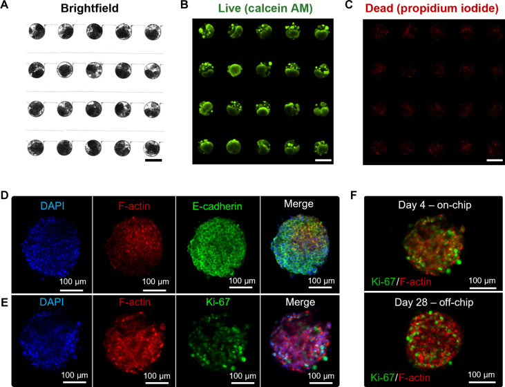
Combination therapies enhance the therapeutic effect of cancer treatment; however, identifying effective interdependent doses, durations, and sequences of multidrug administration regimens is a time- and labor-intensive task. Here, we integrated machine learning, automation, and large microfluidic arrays of cancer spheroids or patient-derived organoids formed in a tissue-mimetic hydrogel to achieve notable acceleration of the discovery of effective multidrug administration regimens. For the clinically approved drug combination, we found a sequential administration regimen leading to a substantial reduction in the total drug dose, in comparison with concurrent drug supply, both at comparable drug efficacy. For the drugs that are currently under clinical development, we found a synergistic effect of concurrently administered drugs and showed that the synergy diminishes for the sequential drug supply. The developed strategy holds promise for the discovery of effective combination therapies for advanced cancer treatment, including personalized chemotherapies.


Figures

Fig. 1.
Fig. 1.. Closed-loop MF tumor-on-a-chip platform integrated with ML.
(A) Schematics of the closed-loop workflow for drug screening. (B) Design of the quadruplet unit with 25 microwells in each row. Scale bar, 1 mm. (C) MF device containing 12 quadruplets. Scale bar, 1 cm. (D) Schematics of the automated fluidic system for sequential administration of multidrug combinations. PDO, patient-derived organoid.
Fig. 2.
Fig. 2.. Growth of MCF-7 spheroids in the MF platform.
(A) Fragment of the array of 300-μm-diameter MCF-7 cell–laden droplets. (B) Fluorescence images of spheroids stained with calcein-AM (green, live cells) and PI (red, dead cells), taken 24 hours after cell loading in the MF device. (C) Immunofluorescence staining of spheroids after 24-hour cell culture using 4′,6-diamidino-2-phenylindole (DAPI) (blue, nuclei), Alexa Fluor 488 E-cadherin rabbit monoclonal antibody (green, cell-cell junctions) and Alexa Fluor 568 phalloidin (red, cytoskeleton). (D) Spheroid staining with Ki-67 rabbit monoclonal antibody (green, proliferative cells), Alexa Fluor 568 phalloidin (red, cytoskeleton), and DAPI (blue, nuclei). The bottom rows in (C) and (D) show merged confocal fluorescence microscopy images.
Fig. 3.
Fig. 3.. Sequential drug administration to MCF-7 spheroids.
(A) Illustration of sequential drug administration. (B) Variation of cell viability, CVexp, achieved in different experimental series [generations (GSs)]. The horizontal red line indicates the optimization target for CVexp. The lowest CVexp achieved for G3-2 simultaneous drug administration is marked with a horizontal cyan line. The most effective sequential schedule is marked with a purple diamond. The data are shown as means ± SD based on three to five independent experiments. (C) Examined sequential drug schedules, organized from left-to-right in the order of ascending CVexp. The color coding indicates the first administered drug, represented by red, blue, and green for DOX, CPA, and 5-FU, respectively. The data are shown as means ± SD based on three to five independent experiments. (D) Validation of the best-performing drug combination administered concurrently and sequentially. Two-sample t test with Holm correction. The data are shown as means ± SD based on more than five independent experiments.
Fig. 4.
Fig. 4.. Validation of sequential drug administration to MCF-7 spheroids.
Comparison of CVexp in the best-performing (A) simultaneous and (B) sequential multidrug administration and individual drug administration [1.25 μM DOX (8 and 48 hours), 105 μM 5-FU (32 and 48 hours), and 94 μM CPA (8 and 48 hours)]. Two-sample t test comparison to the control group with Holm correction. Bars, average of at least, three biological replicates. Error bars, SD. (C) Left to right: Confocal fluorescence microscopy images of spheroids stained with Ki-67 rabbit monoclonal antibody (green, proliferative cells) and with Alexa Fluor 568 phalloidin (red, cytoskeleton); with no treatment (control) and with individual treatment with DOX for 48 and 8 hours, 5-FU for 32 and 48 hours, and CPA for 8 and 48 hours; with concurrent (G3-7) and sequential (GS2-7) administration of three-drug combination. Scale bars, 50 μm. (D) Flow cytometry profiles for MCF-7 cells stained with Ki-67 antibody (proliferative cells), obtained by spheroid dissociation after drug treatment [as in (A) and (B)]. The lines and the numbers show the fraction of proliferative cells.
Fig. 5.
Fig. 5.. Growth of PDOs in the MF platform.
(A to C) Bright-field (A) and fluorescence images of PDOs stained with calcein-AM (green, live cells) (B) and PI (red, dead cells) (C) 96-hour postseeding. Scale bars, 300 μm. (D) Immunofluorescence staining of PDOs after 96-hour cell culture with Ki-67 rabbit monoclonal antibody (green, proliferative cells), Alexa Fluor 568 phalloidin (red, cytoskeleton), and DAPI (blue, nuclei). (E) Immunofluorescence staining of PDOs with DAPI (blue, nuclei), Alexa Fluor 488 E-cadherin rabbit monoclonal antibody (green, cell-cell junctions), and Alexa Fluor 568 phalloidin (red, cytoskeleton) after 96-hour cell culture. (F) Immunofluorescence staining of PDOs grown 4 days on-chip and 28 days off-chip with Ki-67 rabbit monoclonal antibody (green, proliferative cells) and Alexa Fluor 568 phalloidin (red, cytoskeleton).
Fig. 6.
Fig. 6.. Sequential drug administration to PDOs.
(A) Illustration of sequential drug administration. (B) Variation of cell viability, CVexp, in different experimental series (GSs). The horizontal red line indicates the optimization target. The most effective drug combination identified in the GS-2 generation is marked with a purple diamond. The data are shown as means ± SD based on three to five independent experiments. (C) Drug schedules, sorted from left to right in the order of ascending CVexp. The color (red or blue) represents the first administered drug (OLA or IBET-762, respectively). The data are shown as means ± SD based on three to five independent experiments. (D) Comparison of CVexp values for the best-performing simultaneous drug combination G3-2 with sequentially administered multidrug combination GS2-2. Two-sample t test with Holm correction. The data are shown as means ± SD based on more than four independent experiments.
Fig. 7.
Fig. 7.. Validation of sequential drug administration to PDOs.
Comparison of CVexp for best-performing (A) concurrent and (B) sequential administered multidrug combination with the results of PDO treatment with individual drugs [116 μM OLA (96 and 30 hours) and 0.993 μM IBET-762 (96 and 66 hours)]. Two-sample t test with Holm correction). The data are shown as means ± SD based on three to five independent experiments. (C) Left to right: Confocal fluorescence microscopy images of PDOs stained with H2AX rabbit monoclonal antibody (green, DBSs in DNA) and Alexa Fluor 568 phalloidin (red, cytoskeleton); with no treatment (control) and individual treatment with OLA for 96 and 30 hours and IBET-762 for 96 and 66 hours; and with concurrent (G3-2) and sequential (GS2-2) administration of two drugs. Scale bars, 50 μm. (D) Flow cytometry profiles for cells stained with Ki-67 antibody (proliferative cells), obtained by dissociating PDOs postdrug treatment [as in (A) and (B)]. The line designates the fraction of proliferative cells.

Similar articles

References

    1. Turashvili G., Brogi E., Tumor heterogeneity in breast cancer. Front. Med. 4, 227 (2017). - PMC - PubMed
    1. Harbeck N., Penault-Llorca F., Cortes J., Gnant M., Houssami N., Poortmans P., Ruddy K., Tsang J., Cardoso F., Breast cancer. Nat. Rev. Dis. Primers 5, 66 (2019). - PubMed
    1. Lüönd F., Tiede S., Christofori G., Breast cancer as an example of tumour heterogeneity and tumour cell plasticity during malignant progression. Br. J. Cancer 125, 164–175 (2021). - PMC - PubMed
    1. Telli M. L., Carlson R. W., First-line chemotherapy for metastatic breast cancer. Clin. Breast Cancer 9, S66–S72 (2009). - PubMed
    1. Hortobagyi G. N., Buzdar A. U., Current status of adjuvant systemic therapy for primary breast cancer: Progress and controversy. CA Cancer J. Clin. 45, 199–226 (1995). - PubMed