. 2025 Jul 30;17(809):eadw0721.
doi: 10.1126/scitranslmed.adw0721. Epub 2025 Jul 30.

Vaccination with an mRNA-encoded membrane-bound HIV envelope trimer induces neutralizing antibodies in animal models

Affiliations

Vaccination with an mRNA-encoded membrane-bound HIV envelope trimer induces neutralizing antibodies in animal models

Parham Ramezani-Rad et al. Sci Transl Med. .

Abstract

A protective vaccine against human immunodeficiency virus (HIV) will likely need to induce broadly neutralizing antibodies (bnAbs) that engage relatively conserved epitopes on the HIV envelope glycoprotein (Env) trimer. Nearly all vaccine strategies to induce bnAbs require the use of complex immunization regimens involving a series of different immunogens, most of which are Env trimers. Producing protein-based clinical material to evaluate such relatively complex regimens in humans presents major challenges in cost and time. Furthermore, immunization with HIV trimers as soluble proteins induces strong nonneutralizing responses to the trimer base, which is normally occluded on the virion. These base responses could potentially detract from the elicitation of nAbs and the eventual induction of bnAbs. mRNA vaccine platforms offer potential advantages over protein delivery for HIV vaccine development, including increased production speed, reduced cost, and the ability to deliver membrane-bound trimers that might facilitate improved immuno-focusing to nonbase epitopes. We report the design of mRNA-delivered soluble and membrane-bound forms of a stabilized native-like Env trimer (BG505 MD39.3); initial immunogenicity evaluation in rabbits that triggered clinical evaluation; and more comprehensive evaluation of B cell, T cell, and antibody responses in nonhuman primates. mRNA-encoded membrane-bound Env immunization elicited reduced off-target base-directed Env responses and stronger nAb responses compared with mRNA-encoded soluble Env. Overall, mRNA delivery of membrane-bound Env appears promising for enhancing B cell responses to subdominant epitopes and facilitating rapid translation to clinical testing, which should assist HIV vaccine development.


Conflict of interest statement

Competing interests: J.M.S., S.M., and W.R.S. are inventors on a patent for the BG505 MD39 immunogen (US20240374731A1). C.A.C., A.L., S.M., T.S., J.M.S and W.R.S. are inventors on a patent for the BG505 MD39.3 gp151 immunogen (Modified immunogenic proteins, US20230190914A1). S.H. and W.R.S. are employees and shareholders of Moderna, Inc. C.A.C. is the founder and director of the Vaccine Research Foundation. D.J.I. and S.C. are inventors on a patent for the SMNP adjuvant (US11547672B2). All other authors declare no competing interests.

Figures

Fig. 1.
Fig. 1.. Membrane-bound Env mRNA elicited lower base-directed antibody responses than soluble Env in rabbits.
(A) Illustration of MD39.3 mRNA immunogen expression. The cartoon illustrates the in vivo translation of MD39.3 mRNA in transfected cells, producing either soluble or membrane-bound MD39.3 proteins. All MD39.2 and MD39.3 variants are designed with furin cleavage independence provided by a Link14 linker. MD39.3 has a filled glycan hole achieved through N241/N289 glycans. The red circle denotes a mutation in the CD4 binding site to block binding to CD4. (B) ED50 values for serum antibodies binding to matched parental antigens or their BaseKO versions were measured by ELISA. Sera from the gp120 foldon control group were tested against matched antigen and MD39.2 or MD39.3 antigens. Statistical comparisons were performed within each group between antigens. (C) Shown are the ratios of AUC values for BaseKO antigen over AUC values for parental antigen. Lower values indicate the presence of more base binding antibodies in the sera. Statistical comparisons were performed between soluble and membrane-bound forms of MD39.2 and MD39.3. (D) Serum neutralization against BG505 T332N pseudovirus is reported as ID50 values. Statistical comparisons were performed between soluble and membrane-bound forms of MD39.2 and MD39.3. Bars indicate median values for ED50, AUC measurements, and neutralization data. Each point indicates a single animal (n=6 per group). Statistical significance was assessed using the Mann-Whitney test or Kruskal-Wallis test, followed by Dunn’s multiple comparisons test. Significance levels are indicated as ns P > 0.05, *P < 0.05, and ***P < 0.001.
Fig. 2.
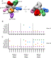
Fig. 2.. Electron microscopy polyclonal epitope mapping characterized immune responses in rabbit serum.
(A) Composite 3D map representing the epitopes observed in negative stain EMPEM analysis. (B and C) Graphs show the number of animals at week 10 (B) and week 26 (C) with detectable antibodies directed to each specific epitope at 10 and 24 weeks post first immunization. Colored to match (A).
Fig. 3.
Fig. 3.. Membrane-bound Env mRNA elicited neutralizing responses in NHPs.
(A) Five groups of six animals per group were immunized with three doses of BG505 MD39.3 immunogens at weeks 0, 8, and 24. All doses were administered bilaterally with each dose split equally between both sides and delivered intramuscularly into the deltoid muscles. Groups 1 to 4 (G1 to G4) received BG505 MD39.3 mRNA immunogens; Group 5 (G5) received BG505 MD39.3 protein plus SMNP adjuvant. The mRNA groups included: soluble MD39.3 (G1), membrane-bound MD39.3 (G2 & G4) and membrane-bound MD39.3 CD4KO (G3). PBMCs were isolated from whole blood and LN FNA from axillary LNs at the indicated time points. (B) Longitudinal ELISA ED50 responses are shown for serum antibodies binding to soluble BG505 MD39.3 gp140. Each point is geometric mean ± geometric standard deviation. (C) Shown are ratios of AUC values for BG505 MD39.3 gp140 BaseKO antigen over AUC values for BG505 MD39.3 gp140 WT. Lower values indicate the presence of more base binding antibodies in the sera. (D) Serum neutralization against BG505 T332N pseudovirus was measured in samples collected at week 26. The frequency of responders is shown below each dataset. Bars indicate median values for AUC measurements and neutralization data. Each point indicates a single animal (n=6 per group) in (C) and (D). Groups 1 and 2 were compared for statistical significance in (C) and (D) using the Mann-Whitney test. Significance levels are indicated as **P < 0.01.
Fig. 4.
Fig. 4.. Electron microscopy polyclonal epitope mapping revealed diverse antibody binding modes elicited by membrane-bound MD39.3 mRNA in NHPs.
(A) Composite 3D map representing the epitopes observed in negative stain EMPEM analysis. (B) Graph showing animals with detectable antibodies against each specific epitope at 26 weeks. Circles on the graph indicate antibodies to the epitope were detected. ID50 values are listed above the graph; nn, no neutralization. Each animal is indicated on the x-axis. Colored to match (A).
Fig. 5.
Fig. 5.. Membrane-bound MD39.3 mRNA induced substantial B cell responses in NHPs.
(A) Representative flow plots show gating for CD20+IgDIgMEnv+/+ BMem cells (left) and BaseKO+ within Env+/+ (right). AF, Alexa Fluor; BV, brilliant violet; PE, phycoerythrin. (B) Frequency of Env+/+ BMem cells of total B cells in PBMCs at different time points are shown. Responses that are lower than the limit of detection (LOD, 0.001375) were set at the LOD. (C) Fold-change of Env+/+ BMem cells per group and time points over baseline Env+/+ BMem frequencies (pre-immunization week −2) is shown. (D) Median frequencies of Env+/+ BMem cells shown in (B) were graphed over time including one additional time point at week 24 (G2 & G5 only). Arrowheads indicate weeks of vaccination. (E) Shown are the percentages of BaseKO+ Env+/+ BMem cells within total Env+/+ BMem cells in all groups post-boosts. G2 values were compared with corresponding G1 values for statistical significance. (F) Shown are the numbers of Env-specific IgG+ BPC in bone marrow (BM) aspirates at week 119 (G1 & G5 only). Bars indicate median for B cell frequencies and each point indicates a single animal (n=6 per group) except (D), which shows median of the respective group. Animals with missing baseline samples were excluded from the fold-change analysis in (C). Groups 1 and 2 were compared for statistical significance using the Mann-Whitney test, followed by the Holm-Šídák multiple comparisons test. Significance levels are indicated as **P < 0.01.
Fig. 6.
Fig. 6.. Membrane-bound MD39.3 mRNA induced both CD4+ and CD8+ T cell responses in NHPs.
(A) Representative flow cytometry plots show gating for Env-specific AIM+ CD4+ T cell responses after stimulation with control DMSO or with MD39.3 peptides; BB, brilliant blue. (B) Env-specific AIM+ CD4+ T cell responses are shown for G2 and G5 at week 2 (post-prime), week 26 (post-third dose) and week −2 (pre-immunization). (C) Representative flow cytometry plots show gating for Env-specific AIM+ CD8+ T cell responses after stimulation with control DMSO or with MD39.3 peptides; Cy, cyanine. (D) Env-specific AIM+ CD8+ T cell responses are shown for G2 and G5 at week 2 (post-prime), week 26 (post-third dose) and week −2 (pre-immunization). Data are shown as background subtracted. Non-responder samples were set at baseline. The dotted black line indicates the limit of quantification (LOQ). Bars represent geometric mean and each point indicate a single animal (n=6 per group, except for G2 which had n = 5 at week 2 or n = 4 at week 26). Pre-immunization had animals from both G2 and G5 (n = 12). The number of samples, the geometric mean (Geo Mean) and the frequency of responders are shown below the plots in (B) and (D). Animals with missing samples or samples exhibiting poor viability were excluded from the analysis. Statistical significance was assessed using the Kruskal-Wallis test, followed by Dunn’s multiple comparisons test. Significance levels are indicated as *P < 0.05, **P < 0.01, and ****P < 0.0001.
Fig. 7.
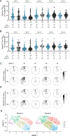
Fig. 7.. Single cell analysis of MD39.3-binding BMem cells indicated somatic hypermutation in NHPs.
(A) Heavy chain mutations in total Env-binding BMem cells were assessed for G2 and G5 at week 8, 10, 24, 26 and 32. (B) Heavy chain mutations on non-base-Env-binding BMem cells (BaseKO+) were assessed for the same groups and timepoints as in (A). Bars indicate median for mutational analysis and each point indicates a single B cell sequence. The number of B cell sequences analyzed, the median number of HC mutations, and the frequency of mutated HCs are shown below each plot. (C) Total Env-binding BMem clonal families were analyzed for groups 2 and 5 across timepoints. Numbers within the Donut plot indicate total number of clonal families detected for each measurement. (D) Non-base-Env-binding BMem clonal families were analyzed for the groups and timepoints as shown in (C). (E) Uniform manifold approximation and projection (UMAP) visualization of single-cell gene expression profiles identifying clusters among Env-binding BMem cells sorted from PBMCs for G2 and G5.

Update of

References

    1. https://www.unaids.org/en/resources/fact-sheet. (2024).
    1. Govindan R, Stephenson KE, HIV Vaccine Development at a Crossroads: New B and T Cell Approaches. Vaccines (Basel) 12, (2024); published online EpubSep 12 ( 10.3390/vaccines12091043). - DOI - PMC - PubMed
    1. Lee JH, Crotty S, HIV vaccinology: 2021 update. Semin Immunol 51, 101470 (2021); published online EpubJan ( 10.1016/j.smim.2021.101470). - DOI - PMC - PubMed
    1. Steichen JM, Phung I, Salcedo E, Ozorowski G, Willis JR, Baboo S, Liguori A, Cottrell CA, Torres JL, Madden PJ, Ma KM, Sutton HJ, Lee JH, Kalyuzhniy O, Allen JD, Rodriguez OL, Adachi Y, Mullen TM, Georgeson E, Kubitz M, Burns A, Barman S, Mopuri R, Metz A, Altheide TK, Diedrich JK, Saha S, Shields K, Schultze SE, Smith ML, Schiffner T, Burton DR, Watson CT, Bosinger SE, Crispin M, Yates JR 3rd, Paulson JC, Ward AB, Sok D, Crotty S, Schief WR, Vaccine priming of rare HIV broadly neutralizing antibody precursors in nonhuman primates. Science 384, eadj8321 (2024); published online EpubMay 17 ( 10.1126/science.adj8321). - DOI - PMC - PubMed
    1. Caniels TG, Medina-Ramirez M, Zhang S, Kratochvil S, Xian Y, Koo JH, Derking R, Samsel J, van Schooten J, Pecetta S, Lamperti E, Yuan M, Carrasco MR, Del Moral Sanchez I, Allen JD, Bouhuijs JH, Yasmeen A, Ketas TJ, Snitselaar JL, Bijl TPL, Martin IC, Torres JL, Cupo A, Shirreff L, Rogers K, Mason RD, Roederer M, Greene KM, Gao H, Silva CM, Baken IJL, Tian M, Alt FW, Pulendran B, Seaman MS, Crispin M, van Gils MJ, Montefiori DC, McDermott AB, Villinger FJ, Koup RA, Moore JP, Klasse PJ, Ozorowski G, Batista FD, Wilson IA, Ward AB, Sanders RW, Germline-targeting HIV vaccination induces neutralizing antibodies to the CD4 binding site. Sci Immunol 9, eadk9550 (2024); published online EpubAug 30 ( 10.1126/sciimmunol.adk9550). - DOI - PMC - PubMed

MeSH terms