Clinical Trial
. 2025 Jul 30;17(809):eady6831.
doi: 10.1126/scitranslmed.ady6831. Epub 2025 Jul 30.

Vaccination with mRNA-encoded membrane-anchored HIV envelope trimers elicited tier 2 neutralizing antibodies in a phase 1 clinical trial

Affiliations
Clinical Trial

Vaccination with mRNA-encoded membrane-anchored HIV envelope trimers elicited tier 2 neutralizing antibodies in a phase 1 clinical trial

K Rachael Parks et al. Sci Transl Med. .

Abstract

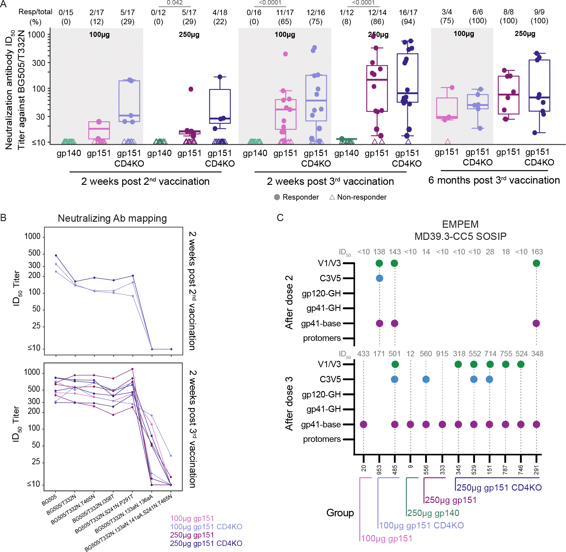
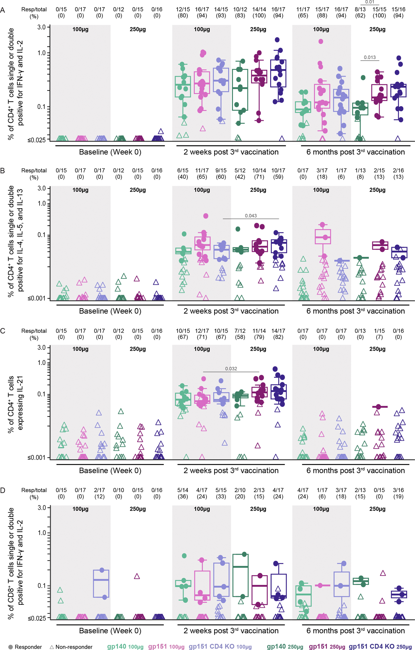
mRNA technology might accelerate development of an urgently needed preventive human immunodeficiency virus (HIV) vaccine. We evaluated the safety and immunogenicity of three mRNA-encoded envelope trimers, including two doses of soluble and membrane-anchored forms, in a randomized, open-label, phase 1 clinical trial. Vaccines were generally well tolerated, although 6.5% (7 of 108) of participants developed urticaria, a higher proportion than seen with other mRNA vaccines. mRNA-encoded trimers induced strong envelope-specific B and T cell responses. Immunization with membrane-anchored trimers, intended to obscure epitopes at the trimer base targeted by nonneutralizing antibodies, reduced the frequency of base-binding serum antibodies in comparison with soluble trimers. Three immunizations elicited autologous tier 2 serum neutralizing antibodies in 80% of vaccinees receiving the membrane-anchored trimers, in contrast to only 4% receiving the soluble trimer. Thus, with demonstration of more favorable safety, mRNA-encoded membrane-anchored HIV envelope trimers represent a promising platform for HIV vaccine clinical development.


Conflict of interest statement

Competing interests: W.R.S. is an inventor on a patent for the BG505 MD39 immunogen (Immunogenic trimers, US20240374731A1). C.A.C., A.L., and W.R.S. are inventors on a patent for the BG505 MD39.3 gp151 immunogen (Modified immunogenic proteins, US20230190914A1). C.A.C. is the founder and director of the Vaccine Research Foundation. SRW has received institutional funding from the National Institute of Allergy and Infectious Diseases/National Institutes of Health; and institutional grants or contracts from Sanofi Pasteur, Janssen Vaccines/Johnson & Johnson, AbbVie, Moderna, Pfizer, Vir Biotechnology, and Worcester HIV Vaccine; has participated on data safety monitoring or advisory boards for Janssen Vaccines/Johnson & Johnson and BioNTech; and his spouse holds stock/stock options in Regeneron Pharmaceuticals. KWC, SH, BL, CR, and WRS are employees of Moderna and may hold stock/stock options in the company. SAR has received institutional funding from the National Institute of Allergy and Infectious Diseases/National Institutes of Health; and institutional grants or contracts from Merck and Gilead Sciences. MJM receives institutional funding from the National Institute of Allergy and Infectious Diseases/National Institutes of Health, and she has received institutional grants or contracts from Bill and Melinda Gates Foundation, IAVI, Sanofi Pasteur, Moderna, and Regeneron Pharmaceuticals.

Figures

Figure 1.
Figure 1.. Trial schema.
(A) Graphic depiction of the HVTN 302 study groups and vaccine doses in Parts A and B, vaccine delivery schedule and enrollment strategy for safety holds. For planned safety hold #1, a safety sentinel cohort of participants in Part A (n=4 each group, 12 total) were enrolled and evaluated for safety for 2 weeks after the first vaccination. If safety criteria were met, the remaining Part A participants were enrolled, as well as a subset of participants from Part B. For planned safety hold #2, a safety sentinel cohort of participants in Part B (n=4 each group, 12 total) were enrolled and evaluated for safety for 2 weeks after the first vaccination. If safety criteria were met, the remaining Part B participants were enrolled. (B) Time points of blood sample collection for immune analyses.
Figure 2.
Figure 2.. Vaccine-specific serum binding antibody responses were elicited by vaccination.
(A and B) IgG binding antibody responses to BG505 MD39.3 (A) and BG505 MD39.3 (B) base epitopes were measured by BAMA and titers reported as area under the curve (AUC) for the different vaccine groups, specified by color-coding defined below (B). Positive response rates and magnitudes among positive responders to BG505 MD39.3 are shown at 2 weeks after the 1st, 2nd, and 3rd vaccinations, and at 6 months after 3rd vaccination. The proportion of positive responders is shown by vaccine group at the top of (A). Boxplots display the distribution of positive responses across participants by vaccine group with medians indicated by mid-lines; the ends of the boxes denote the 25th and 75th percentiles and the whiskers extend to the most extreme data points that are no more than 1.5 times the interquartile range (the height of the box) or if no value meets this criterion, to the data extremes. Circles denote responders; triangles represent non-responders. Significant p-values are shown for comparisons of response rates by Barnard’s test (listed above Resp/total) and magnitudes among positive responders by Wilcoxon test (listed above boxplots) for the following group comparisons: gp140 vs. gp151 at each dose, gp151 vs. gp151 CD4KO at each dose, gp140 100μg vs. gp140 250μg, gp151 100μg vs. gp151 250μg, gp151 CD4KO 100μg vs. gp151 CD4KO 250μg.
Figure 3.
Figure 3.. Neutralizing antibody responses (autologous and glycan mutant panel) and EMPEM responses to vaccination targeted multiple HIV Env epitopes.
(A) Neutralizing antibody ID50 titers against BG505/T33N were measured as a function of reductions in Tat-regulated luciferase (Luc) reporter gene expression in TZM-bl cells for all participants in each vaccine group. Positive response rates and magnitudes among positive responders against BG505/T33N are shown at 2 weeks after 2nd and 3rd vaccinations, and at 6 months after 3rd vaccination for the 27 participants with month 6.5 ID50 ≥ 60 to BG505/T332N. The proportion of positive responders for each vaccine group is shown at the top of (A) where boxplots show the distribution of positive responses across participants in a given vaccine group with medians indicated by mid-lines; the ends of the boxes denote the 25th and 75th percentiles and the whiskers extend to the most extreme data points that are no more than 1.5 times the interquartile range (the height of the box) or if no value meets this criterion, to the data extremes. Circles denote responders; triangles represent non-responders. Significant p-values are shown for comparisons of response rates by Barnard’s test (listed above Resp/total) with no significant differences in magnitudes among positive responders seen by Wilcoxon test for the following group comparisons: gp140 vs. gp151 at each dose, gp151 vs. gp151 CD4KO at each dose, gp140 100μg vs. gp140 250μg, gp151 100μg vs. gp151 250μg, gp151 CD4KO 100μg vs. gp151 CD4KO 250μg. (B) Any samples resulting in >200 ID50 titer against BG505/T332N at 2 weeks after 2nd (n = 3) or 3rd vaccination (n = 10) were tested against a panel of mutant BG505 viruses. The line color denotes vaccine group. (C) Serum from select participants with neutralizing ID50 values to BG505/T332N were assayed by EMPEM 2 weeks following 2nd vaccination or two weeks following 3rd vaccination or both. Dotted gray lines indicate the sample was assayed. Dots on the graph indicate that reactivity to that epitope was detected. ID50 values are listed above the graph. The group of each participant is indicated on the x-axis.
Figure 4.
Figure 4.. BG505 MD39.3-specific memory B cells increased in frequency and somatic hypermutation rate and underwent clonal expansion following vaccination.
(A and B) Frequencies of BG505 MD39.3++ (A) and non-base binding (B) B cells of total IgG+ B cells measured at baseline, 2 weeks post 2nd vaccination, 2 weeks post 3rd vaccination, and 6 months post 3rd vaccination. Each data point for a single participant are shown and colored by vaccine group. Circles denote responders; triangles represent non-responders. The proportion of positive responders by vaccine group is shown at the top of each panel. Significant p-values are shown for comparisons of magnitudes among positive responders by Wilcoxon test (listed above boxplots), with no significant differences in response rates by Barnard’s test for the following group comparisons: gp140 vs. gp151 at each dose, gp151 vs. gp151 CD4KO at each dose, gp140 100μg vs. gp140 250μg, gp151 100μg vs. gp151 250μg, gp151 CD4KO 100μg vs. gp151 CD4KO 250μg. (C) Rates of nucleotide (NT) SHM in V segments of heavy, lambda, and kappa chains for the base- and non-base-specific memory (IgD) B cell receptors at week 0, week 10 and week 26 after vaccination (vacc.). Each dot represents the median rate of SHM across sequenced B cell receptors from one participant. The size of the dot represents the number of sequences recovered, and dots are colored by group. Statistical comparisons of SHM between base and non-base binders at a given time point, and between time points within base or non-base binders, were performed using a Wilcoxon signed-rank test (two-sided) to account for paired data. In panels (A to C), boxplots show the distribution of responses across participants in a given vaccine group with medians indicated by mid-lines; the ends of the box denote the 25th and 75th percentiles and the whiskers extend to the most extreme data points that are no more than 1.5 times the interquartile range (the height of the box) or if no value meets this criterion, to the data extremes. (D) Number of base- and non-base-specific clonotypes with a given size (number of cells) at week 0, week 10, and week 26 within each vaccine group. Lines are colored by group.
Figure 5.
Figure 5.. CD4+ and CD8+ T cell responses to total Env increased after vaccination.
(A to D) CD4+ (A to C) and CD8+ (D) T cell responses were assessed by intracellular cytokine staining. Data are expressed as the percentage of cells producing one or both IFN-γ and IL-2 (A, D); IL-4, IL-5, or IL-13 (B); and IL-21 (C) in the different vaccine groups. CD4+ and CD8+ T cell positive response rates and magnitudes among positive responders to total Env are shown at week 0, 2 weeks after the 3rd vaccination, as well as 6 months after the 3rd vaccination. The proportion of positive responders by group is shown above each panel. Boxplots show the distribution of positive responses in a given vaccine group with medians indicated by mid-lines; the ends of the boxes denote the 25th and 75th percentiles and the whiskers extend to the most extreme data points that are no more than 1.5 times the interquartile range (the height of the box) or if no value meets this criterion, to the data extremes. Circles denote responders; triangles represent non-responders. Significant p-values are shown for comparisons of response rates by Barnard’s test (listed above Resp/total) and magnitudes among positive responders by Wilcoxon test (listed above boxplots) for the following group comparisons: gp140 vs. gp151 at each dose, gp151 vs. gp151 CD4KO at each dose, gp140 100μg vs. gp140 250μg, gp151 100μg vs. gp151 250μg, gp151 CD4KO 100μg vs. gp151 CD4KO 250μg.

References

    1. Beyrer C, Tomaras GD, Gelderblom HC, Gray GE, Janes HE, Bekker L-G, Millett G, Pantaleo G, Buchbinder S, Corey L, Is HIV epidemic control by 2030 realistic? Lancet HIV 11, e489–e494 (2024). - PMC - PubMed
    1. Gray GE, Mngadi K, Lavreys L, Nijs S, Gilbert PB, Hural J, Hyrien O, Juraska M, Luedtke A, Mann P, McElrath MJ, Odhiambo JA, Stieh DJ, van Duijn J, Takalani AN, Willems W, Tapley A, Tomaras GD, Hoof JV, Schuitemaker H, Swann E, Barouch DH, Kublin JG, Corey L, Pau MG, Buchbinder S, Tomaka F, I. 705/HPX2008 S. Group, Allagappen J, Andriesen J, Ayres A, Bekker L-G, Borremans C, Brumskine W, Chilengi R, Dubula T, Garrett N, Gelderblom H, Gill K, Hoosain Z, Hosseinipour M, Hutter J, Inambao M, Innes C, Kilembe W, Kotze P, Kotze S, Laher F, Laszlo I, Lazarus E, Malahleha M, Mathebula M, Matoga M, McClennen R, Mda P, Meerts P, Naicker V, Naidoo L, Philip T, Pitsi A, Scheppler L, Sopher C, Takuva SG, Viegas E, Weijtens M, Yuan O, Mosaic HIV-1 vaccine regimen in southern African women (Imbokodo/HVTN 705/HPX2008): a randomised, double-blind, placebo-controlled, phase 2b trial. Lancet Infect. Dis. (2024), doi: 10.1016/s1473-3099(24)00358-x. - DOI - PMC - PubMed
    1. NIH, Experimental HIV vaccine regimen safe but ineffective, study finds (2023) (available at https://www.nih.gov/news-events/news-releases/experimental-hiv-vaccine-r...).
    1. Leggat DJ, Cohen KW, Willis JR, Fulp WJ, deCamp AC, Kalyuzhniy O, Cottrell CA, Menis S, Finak G, Ballweber-Fleming L, Srikanth A, Plyler JR, Schiffner T, Liguori A, Rahaman F, Lombardo A, Philiponis V, Whaley RE, Seese A, Brand J, Ruppel AM, Hoyland W, Yates NL, Williams LD, Greene K, Gao H, Mahoney CR, Corcoran MM, Cagigi A, Taylor A, Brown DM, Ambrozak DR, Sincomb T, Hu X, Tingle R, Georgeson E, Eskandarzadeh S, Alavi N, Lu D, Mullen T-M, Kubitz M, Groschel B, Maenza J, Kolokythas O, Khati N, Bethony J, Crotty S, Roederer M, Hedestam GBK, Tomaras GD, Montefiori D, Diemert D, Koup RA, Laufer DS, McElrath MJ, McDermott AB, Schief WR, Vaccination induces HIV broadly neutralizing antibody precursors in humans. Science 378, eadd6502 (2022). - PMC - PubMed
    1. Steichen JM, Phung I, Salcedo E, Ozorowski G, Willis JR, Baboo S, Liguori A, Cottrell CA, Torres JL, Madden PJ, Ma KM, Sutton HJ, Lee JH, Kalyuzhniy O, Allen JD, Rodriguez OL, Adachi Y, Mullen T-M, Georgeson E, Kubitz M, Burns A, Barman S, Mopuri R, Metz A, Altheide TK, Diedrich JK, Saha S, Shields K, Schultze SE, Smith ML, Schiffner T, Burton DR, Watson CT, Bosinger SE, Crispin M, YatesIII JR, Paulson JC, Ward AB, Sok D, Crotty S, Schief WR, Vaccine priming of rare HIV broadly neutralizing antibody precursors in nonhuman primates. Science 384, eadj8321 (2024). - PMC - PubMed

Publication types