Review
. 2025 Jul 31;10(1):227.
doi: 10.1038/s41392-025-02280-1.

Immune evasion in cancer: mechanisms and cutting-edge therapeutic approaches

Affiliations
Review

Immune evasion in cancer: mechanisms and cutting-edge therapeutic approaches

Muhammad Tufail et al. Signal Transduct Target Ther. .

Abstract

Immune evasion represents a significant challenge in oncology. It allows tumors to evade immune surveillance and destruction, thereby complicating therapeutic interventions and contributing to suboptimal patient outcomes. This review addresses the critical need to understand how cancers evade immune surveillance. It aims to provide a comprehensive overview of strategies of tumors to escape immune detection by examining tumor-induced immune suppression, immune checkpoint regulation, and genetic and epigenetic influences. Moreover, it explores the dynamic role of the tumor microenvironment (TME) in fostering immune resistance and highlights the impact of metabolic reprogramming on immune suppression. Additionally, this review focuses on how tumor heterogeneity influences immune evasion and discusses the limitations of current immunotherapies. The role of key signaling pathways, including programmed cell death protein 1/programmed cell death ligand 1 (PD-1/PD-L1), cytotoxic T-lymphocyte-associated antigen 4 (CTLA-4), transforming growth factor-β (TGF-β), nuclear factor kappa-light-chain-enhancer of activated B cells (NF-κB), and cyclic GMP-AMP synthase-stimulator of interferon genes (cGAS-STING) is analyzed to elucidate their contributions to immune escape. Emphasizing the complexities of immune evasion, this review underscores the importance of personalized approaches and the integration of multi-omics data to combat therapeutic resistance. Furthermore, it discusses novel and emerging therapeutic strategies, such as bispecific antibodies, oncolytic viruses, and nanotechnology-driven immunotherapies, showcasing innovative avenues in cancer treatment. The significance of this review lies in its potential to guide future research and innovations in immunotherapy, ultimately improving patient outcomes and advancing our understanding of cancer immunology.


Conflict of interest statement

Competing interests: The authors declare no competing interests. Consent for publication: All authors have given consent for publication.

Figures
Fig. 1
Fig. 1
Mechanisms of immune evasion in cancer. This figure illustrates the diverse mechanisms by which cancer cells evade immune surveillance. At the center, the main theme, “Immune Evasion in Cancer,” branches out into four key mechanisms. Tumor-induced immune suppression includes inhibitory cytokines and chemokines, Tregs, and MDSCs, all of which contribute to an immunosuppressive environment. Immune Checkpoint Regulation highlights pathways like PD-1/PD-L1, CTLA-4, and other inhibitory checkpoints, which tumors exploit to deactivate immune cells. In TME modulation, components like hypoxia, metabolic reprogramming, stromal cells, and ECM create physical and biochemical barriers that hinder immune cell infiltration and activation. Antigen presentation and recognition pathways are compromised through MHC class I downregulation, immune editing, etc., reducing the visibility of cancer cells to cytotoxic T cells
Fig. 2
Fig. 2
Immune evasion mechanisms driven by oncogenes and tumor suppressor gene loss in cancer. Oncogenes and tumor suppressor genes contribute to immune evasion through diverse mechanisms, aiding tumor cells in avoiding immune detection and elimination. For example, the Myc increases the expression of PD-L1 and leads to immune suppression. Similarly, the RAS family can increase CD47, which leads to T cell inhibition. Mutations in the EGFR, BRAF, and HER2 upregulate PD-L1 expression, leading to immune evasion. STAT3 promotes immune suppression by inducing PD-L1 and immunosuppressive cytokines. In contrast, tumor suppressor gene loss, such as TP53 and PTEN, increases PD-L1 and PI3K pathway activation to enhance immune evasion. RB1 loss increases immunosuppressive factors, preventing immune cell infiltration. APC loss activates the Wnt/β-catenin pathway, reducing T cell infiltration. BRCA1/2 and SMAD4 loss promote the recruitment of regulatory T cells and MDSCs, strengthening the immunosuppressive microenvironment. ATM loss suppresses immune signaling related to the DNA damage response, further reducing immune activation. Each of these genetic changes fosters immune evasion, creating a hostile microenvironment for anti-tumor immune responses
Fig. 3
Fig. 3
DNA methylation, histone modification, and immune evasion in cancer. This figure illustrates the interplay between key immune-related genes and DNA methylation and histone modification in orchestrating the immune response against tumors in cancer immunology. In the DNA methylation section, hypermethylation of the promoter regions of immune-related genes is shown to repress their expression, reducing immune cell infiltration into the tumor. This is depicted by a DNA strand marked by methyl groups, which connect to reduced gene expression and limited immune presence near the tumor. In the histone modification section, HDACs and histone acetyltransferases (HATs) modify chromatin structure, either condensing it (heterochromatin) or relaxing it (euchromatin), respectively. HDAC activity leads to closed chromatin around immune-regulatory genes, silencing their expression, while HAT activity may open chromatin but does not effectively activate immune-activating genes in tumors. These repressive changes result in immunosuppressive TME, with immune cells depicted as partially blocked or inactive. This illustrates how epigenetic modifications support immune evasion by repressing immune-modulatory molecules and limiting immune cell access to the tumor
Fig. 4
Fig. 4
Tumor heterogeneity and immune evasion in cancer. This figure illustrates intra-tumor heterogeneity and the spatial-temporal dynamics of immune evasion in cancer. The diversity of cell populations in the tumor enables unique immune escape strategies that enhance resistance and contribute to cancer progression. Cancer stem cells exhibit self-renewal and releasing immune-suppressive factors to aid regrowth and metastasis. Mesenchymal-like cells undergo EMT, gaining motility and reducing antigen presentation for immune evasion. Cells expressing PD-L1 inhibit T cell activity through checkpoint interactions, while those with low MHC expression avoid cytotoxic detection by reducing antigen display. Hypoxic cells thrive in low-oxygen zones, fostering an immunosuppressive microenvironment and secret factors that create immune responses in cytokine-producing cells. Highly proliferative, mutationally diverse cells vary in antigen expression, complicating immune targeting, while dormant/quiescent cells evade detection through reduced activity but may later drive recurrence. Spatial heterogeneity within the TME and the variable distribution of immune cell populations across these regions are shown. A timeline tracking clonal evolution from primary tumor to metastasis illustrates temporal heterogeneity throughout tumor progression. There is also the emergence of resistance mechanisms, while depicted immune evasion strategies evolve over time, including loss of antigen presentation and increased regulatory T cells
Fig. 5
Fig. 5
Pathways associated with the immune evasion of cancer. a The PD-1/PD-L1 pathway in immune evasion of cancer illustrates how binding between PD-1 receptors on T cells and PD-L1 on cancer cells triggers inhibitory signaling that suppresses T cell functions. This interaction recruits SHP-2 phosphatase to PD-1’s ITIM and ITSM motifs, which dephosphorylates key signaling molecules, inhibiting the PI3K-Akt, RAS-MAPK, and JAK-STAT pathways. This suppression decreases cytokine production, cytotoxic activity, and T cell proliferation and survival, leading to T cell exhaustion. Together, these effects contribute to immune evasion, tumor growth, and potential metastasis due to impaired immune surveillance in the TME. b CTLA-4 Pathway in immune evasion of cancer shows how CTLA-4 suppresses T cell activation to aid immune evasion. T cell activation typically requires CD28 binding to B7 ligands on APCs. However, CTLA-4, with its higher affinity for B7, outcompetes CD28, leading to reduced T cell activation. This CTLA-4-B7 binding suppresses intracellular signaling pathways in T cells, including the PI3K-Akt and NF-κB pathways, resulting in decreased T cell proliferation, reduced cytokine production, and impaired immune response. The outcome is immune evasion, allowing tumor cells to survive and proliferate. c TGF-β is secreted by various cells, binding to TGF-β receptors on immune cells. This interaction initiates intracellular signaling cascades, particularly through the SMAD pathway, leading to gene transcription that suppresses immune functions. Key effects include reduced production of pro-inflammatory cytokines, diminished cytotoxic molecule release, and a shift in T cell differentiation toward immunosuppressive Tregs, collectively weakening the immune response. d The IL-10 binds to IL-10 receptors. This binding activates the JAK1/STAT3 pathway, leading to the transcription of immunosuppressive genes that reduce immune responses. In CD8+ T cells, IL-10 signaling suppresses cytokine production and cytotoxic molecules, weakening their ability to attack cancer cells. In APCs, IL-10 impedes maturation, decreases antigen presentation, and downregulates co-stimulatory molecules, thereby limiting T cell activation. For NK cells, IL-10 reduces cytotoxic activity and receptor expression, diminishing their tumor-targeting function. IL-10 also promotes Treg differentiation, creating a feedback loop of immunosuppression that supports tumor growth and metastasis. e The NF-κB Pathway in immune evasion of cancer demonstrates how NF-κB activation within both tumor and immune cells promotes immune suppression. In cancer cells, NF-κB signaling induces pro-survival and proliferative genes, along with pro-inflammatory cytokines, fostering a tumor-supportive microenvironment. In immune cells, NF-κB drives the expression of immunosuppressive cytokines, recruiting Tregs and myeloid-derived suppressor cells, which reduce T cell and NK cell activity. Together, these effects enable immune evasion, supporting tumor growth and metastasis. f The figure illustrates the cGAS-STING pathway in immune evasion of cancer, highlighting key molecular and cellular events. Tumor cells release cytosolic DNA due to genomic instability or necrosis, which is sensed by cGAS, leading to the production of cGAMP. cGAMP binds to and activates STING, where it initiates downstream signaling, including the activation of TBK1 and phosphorylation of IRF3. This cascade promotes the production of type I interferons and pro-inflammatory cytokines, which can recruit immune cells. However, STING activation also contributes to immune evasion by inducing an inflammatory microenvironment, the recruitment of immunosuppressive cells, the upregulation of immune checkpoint molecules, and the secretion of immunosuppressive cytokines
Fig. 6
Fig. 6
Therapeutic strategies to overcome immune evasion in cancer. Therapeutic strategies targeting immune evasion in cancer are summarized, highlighting five main approaches: immune checkpoint inhibitors, cancer vaccines, oncolytic viruses, adoptive T cell therapy, and epigenetic modulation. Immune checkpoint inhibitors restore T cell activation by targeting pathways like PD-1/PD-L1 and CTLA-4, but face challenges such as tumor heterogeneity and resistance. Cancer vaccines, including peptide-based and neoantigen vaccines, are often combined with checkpoint inhibitors but are limited by antigen heterogeneity and immunosuppressive mechanisms. Oncolytic viruses promote antigen presentation, cytokine release, and immune activation within the TME, while addressing challenges like antigen escape. Adoptive T cell therapies, including CAR-T cells and tumor-infiltrating lymphocytes, combat issues like antigen loss, MHC downregulation, and immunosuppressive signaling. Epigenetic modulation targets DNA methylation and histone modifications to reverse immune suppression, reprogram the TME, and restore antigen presentation. Together, these approaches tackle the complexities of immune evasion and aim to enhance cancer immunotherapy outcomes
Fig. 7
Fig. 7
Strategies employed by CAR-T cells to counter immune evasion in cancer. This figure illustrates the multifaceted strategies employed by CAR-T cells to overcome immune evasion in cancer. CAR-T cells are engineered for targeted antigen recognition, allowing precise targeting of tumor-specific antigens, thereby minimizing off-target effects. Checkpoint inhibition is achieved by combining CAR-T cells with agents that block immune checkpoints, effectively countering inhibitory signals from the tumor. To resist the TME immunosuppressive influences, CAR-T cells are designed to withstand suppressive factors. Cytokine secretion by CAR-T cells amplifies the immune response by recruiting and activating other immune cells within the tumor vicinity. Additionally, dual-targeting CAR-T cells address tumor heterogeneity by recognizing multiple antigens, thereby reducing the risk of immune evasion through antigen loss. Lastly, armored CAR-T cells are engineered to express additional receptors, enhancing resilience against the hostile TME and supporting sustained immune activity
Fig. 8
Fig. 8
Advantages of nanotechnology and drug delivery systems in cancer treatment. This figure illustrates the advantages of nanotechnology and drug delivery systems in cancer treatment. Nanoparticles enhance targeted drug delivery by selectively binding to cancer cells, minimizing the exposure of healthy tissues, and reducing systemic toxicity. The enhanced permeability and retention (EPR) effect allows nanoparticles to accumulate at tumor sites due to leaky blood vessels, improving drug retention and therapeutic efficacy. Controlled drug release mechanisms ensure sustained and precise drug delivery, while multifunctionality enables simultaneous therapy and monitoring. Additionally, nanoparticles facilitate combination therapies, improve biodistribution by evading immune clearance, and allow for personalized treatment approaches, optimizing patient outcomes and minimizing side effects
Fig. 9
Fig. 9
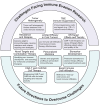
Challenges and future directions in overcoming immune evasion in cancer. This figure outlines the key challenges and future directions in addressing immune evasion in cancer. The “Challenges” section highlights issues such as tumor heterogeneity, where spatial and temporal variations in immune resistance complicate treatment; immune suppression within the TME, driven by cells like Tregs and MDSCs and inhibitory signals like PD-L1 expression; treatment resistance and relapse, which require novel strategies for sustained responses; the limited efficacy of immunotherapies in “cold tumors” with low immune cell infiltration; and off-target effects and toxicity associated with immune-based therapies like CAR-T cells. The “Future Directions” section focuses on potential solutions, including the identification of novel therapeutic targets such as new immune checkpoints and tumor antigens, combination therapies that integrate immune checkpoint inhibitors with other treatments, personalized medicine approaches based on genetic and epigenetic profiling, the development of predictive biomarkers for early assessment of therapy efficacy and resistance, advancements in CAR-T and CAR-NK cell therapies to enhance targeting and reduce toxicity, and strategies to increase immune cell infiltration in cold tumors, such as oncolytic viruses or local modifications of the TME

References

    1. Caner, A. Immune escape mechanism of cancer. Curr. Mol. Biol. Rep.10, 9–19 (2024).
    1. Li, Y. et al. Immune cycle-based strategies for cancer immunotherapy. Adv. Funct. Mater.31, 2107540 (2021).
    1. Dutta, S. et al. Targets of immune escape mechanisms in cancer: basis for development and evolution of cancer immune checkpoint inhibitors. Biology12, 218 (2023). - PMC - PubMed
    1. Messerschmidt, J. L., Prendergast, G. C. & Messerschmidt, G. L. How cancers escape immune destruction and mechanisms of action for the new significantly active immune therapies: helping nonimmunologists decipher recent advances. Oncologist21, 233–243 (2016). - PMC - PubMed
    1. O’Connell, P. Uncovering mechanisms underlying complement-mediated cancer immune evasion. Mol. Ther.32, 277–278 (2024). - PMC - PubMed