. 2025 Jul 30;8(1):1134.
doi: 10.1038/s42003-025-08525-7.

Inhibition of inner ear macrophage phagocytosis alleviates cisplatin-induced ototoxicity

Affiliations

Inhibition of inner ear macrophage phagocytosis alleviates cisplatin-induced ototoxicity

Jiahui Zhang et al. Commun Biol. .

Abstract

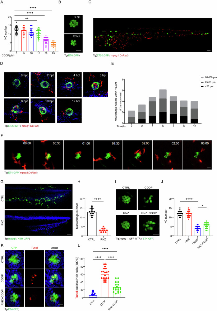
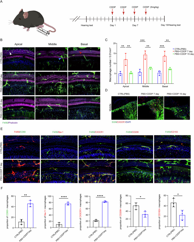
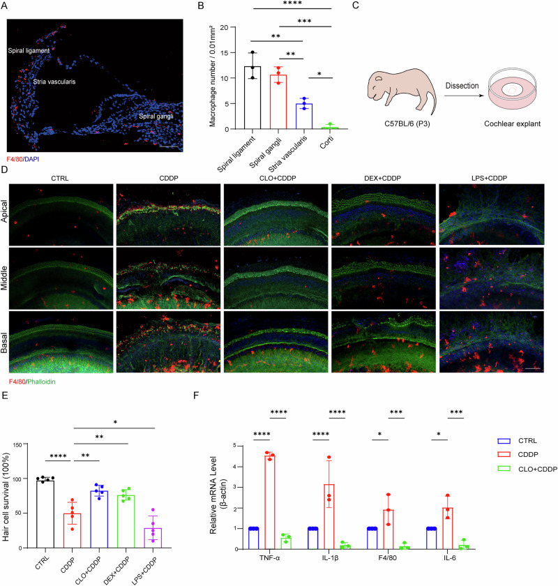
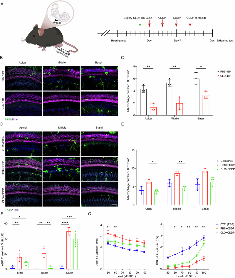
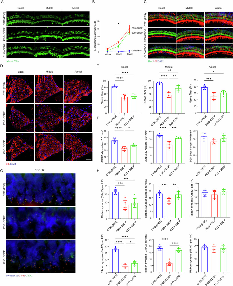
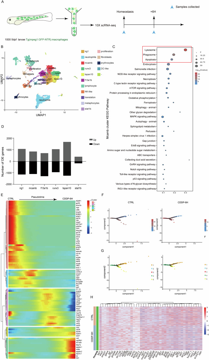
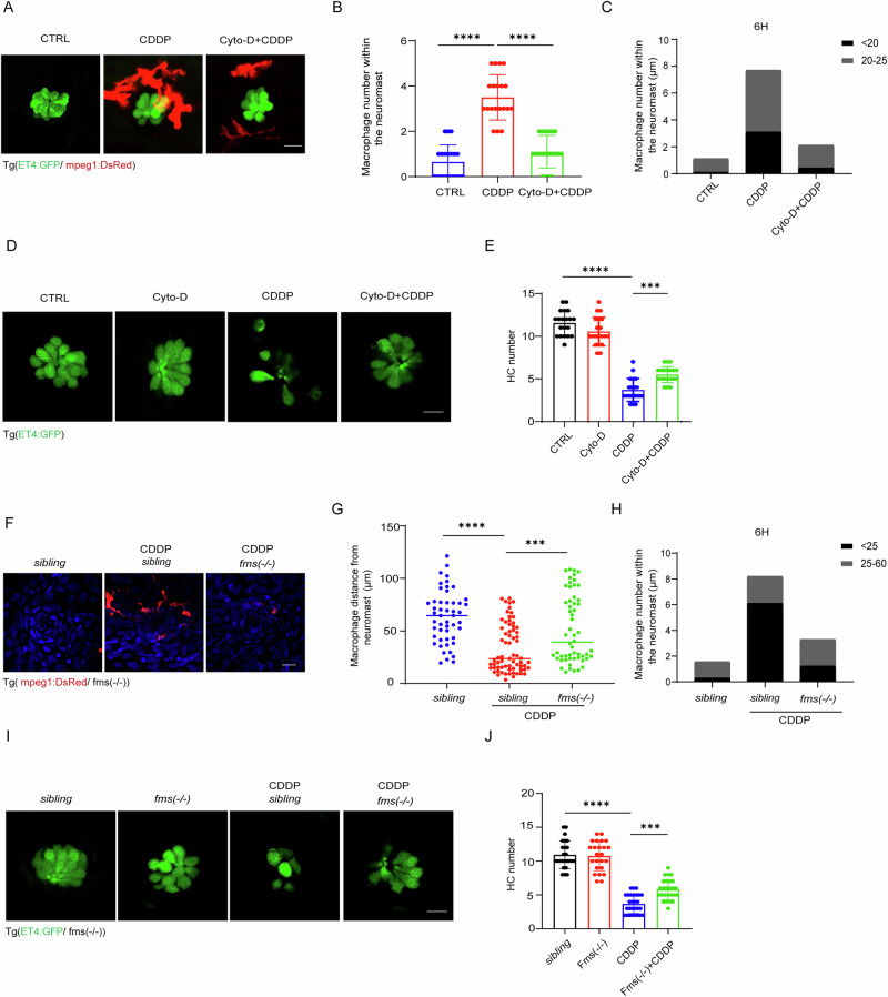
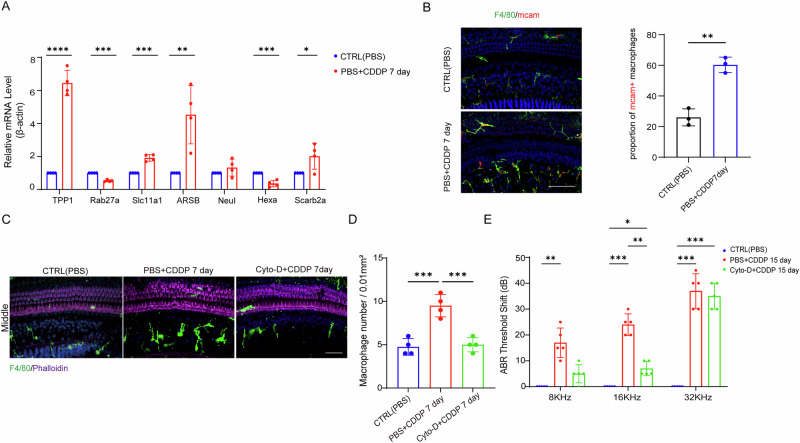
The immune response is considered a significant pathological mechanism of inner ear damage. However, the role of macrophages, as key components of immune cells, in immunity in the inner ear remains elusive. Evidence from other organs indicates that phagocytosis, a core function of macrophages, plays a crucial role in maintaining homeostasis, development, and tissue repair regeneration. However, it has rarely been studied in the inner ear. This field may currently hold new insights. In this study, we aimed to investigate the immunological contribution of resident macrophages in the inner ear to cisplatin-induced ototoxicity. By using clodronate liposomes and cytochalasin to deplete macrophages or inhibit macrophage phagocytosis locally, we first elucidated the dynamic changes in the immune state of inner ear macrophages during cisplatin injury through multimodal and multidimensional approaches. High-spatiotemporal-resolution single-cell analysis and real-time imaging of macrophages during zebrafish hair cell death identified proinflammatory subsets during cisplatin injury. We found that macrophage activation through phagocytosis synergized with the inflammatory response and that inhibiting macrophage phagocytosis could ameliorate cisplatin-induced ototoxicity. Finally, we discuss how the highly plastic phagocytic function of resident macrophages in the inner ear holds potential for the development of strategies for treating cisplatin-induced hearing loss.


Conflict of interest statement

Competing interests: The authors declare no competing interests. Ethical approval: All animal experiments conducted in this study are approved by the Institutional Animal Care and Use Committee (IACUC) of Sun Yat-sen University. We have complied with all relevant ethical regulations for animal use.

Figures

Fig. 1
Fig. 1. In zebrafish, macrophages promote cisplatin-induced hair cell death.
A Changes in the number of hair cells in the lateral line of zebrafish after treatment with different concentrations of cisplatin (0–25 μM) (n = 10 zebrafish for each group). B Hair cell counts decreased in zebrafish larvae after 12 hour post-treatment (hpt) with 25 μM cisplatin, scale bar = 20 μm. C Fluorescence images showing the distribution of neuromasts and macrophages in zebrafish larvae 5-day post-fertilization (mantle cells: green, macrophages: red), scale bar = 100 μm. D and F Dynamics of macrophages (red) residing on the surface of zebrafish approaching-neighboring-engulfing-leaving the neuromasts (green) after cisplatin treatment, scale bar = 20 μm. E The distribution of macrophages within different diameter ranges around the neuromasts was statistically analyzed (n = 8 zebrafish for each group). G 2.5 mM RNZ-treated fluorescence images of macrophages. H Quantification of the effect of macrophage clearance within 500 μm of the tail (n = 10 zebrafish for each group), scale bar = 100 μm. I Changes in the number of hair cells in the zebrafish lateral line system before and after macrophage clearance following cisplatin treatment, scale bar = 20 μm. J Statistical analysis of hair cell survival before and after macrophage clearance (n = 10 zebrafish for each group). K Red fluorescence signals in neuromast hair cells (green) originate from TUNEL staining, specifically indicating apoptotic hair cells, scale bar = 20 μm. L Quantification of the TUNEL-positive hair cells (n = 10 zebrafish for each group). Statistical analyses were carried out via one-way ANOVA for (A) and (L), two-way ANOVA for (J) and t-test for (H). The data were presented as mean ± SD. *p < 0.05, **p < 0.01, ***p < 0.001, ****p < 0.0001.
Fig. 2
Fig. 2. Macrophages are recruited to and activated in the cochlea of mice after cisplatin administration.
A Cisplatin treatment timeline for C57BL/6 mice. Created in BioRender. https://BioRender.com/x92914j. B Representative fluorescence images of macrophages at 0 day, 7 day, 15 day post-cisplatin exposure showing changes in the quantity and morphology distribution of macrophages (Hair cells: purple, macrophages: green). Typical macrophages were indicated with arrows respectively, stellate appearance in the apical (white), dendritic appearance in the middle (blue), and ameboid appearance at the basal (red), scale bar = 20 μm. C Calculations on basilar membrane confocal image stacks, using outer hair cells as the origin, with the direction toward the cochlear modiolus, respectively present the number of macrophages from the apical, middle, and basal turns of the cochlea within each 0.1 mm × 0.1 mm region after cisplatin treatment 7 day and 15 day (n = 3 mice for each group). D Representative fluorescence images of macrophages and spiral ganglion neurons (SGNs) in frozen sections of CX3CR1GFP/+ mice (SGN: green, CX3CR1: red), scale bar = 40 μm. E Representative fluorescence images of different macrophage marker labels (including CD68, Iba-1, CX3CR1, CD206, and CD163), scale bar = 40 μm. F Quantitative analysis of macrophage proportion expressing different markers within the cochlea on the 7-day post-cisplatin treatment (n = 3 mice for each group). Statistical analyses were carried out via two-way ANOVA for (C) and t-test for (F). The data were presented as mean ± SD.*p < 0.05, **p < 0.01, ***p < 0.001, ****p < 0.0001.
Fig. 3
Fig. 3. Eliminating macrophages or inhibiting their inflammatory activation can alleviate CDDP-induced hair cell death in cochlea explants.
A Fluorescence image showing the distribution of macrophages in frozen sections of the cochlea from P3 mice (macrophages: red), scale bar = 50 μm. B Quantitative analysis of macrophages within each 0.1 mm × 0.1 mm region (n = 3 mice for each group). C Cochlea explant culturing schematic. Created in BioRender. https://BioRender.com/d9ejdlc. D Fluorescence images displaying changes of macrophages after cisplatin exposure in cochlea explants (Phalloidin: green, macrophages: red), scale bar = 40 μm. E Quantification of hair cells labeled with phalloidin in (D) (n = 5 mice for each group). F RT‒PCR analysis of F4/80, TNF-α, IL-1β, and IL-6 gene expression levels (n = 3, biologically independent experiments). Statistical analyses were carried out via one-way ANOVA for (B) and (E), and two-way ANOVA for (F). The data were presented as mean ± SD.*p < 0.05, **p < 0.01, ***p < 0.001, ****p < 0.0001.
Fig. 4
Fig. 4. Clearance of resident macrophages reduces cisplatin-induced hearing loss.
A Schematic diagram of intraperitoneal injection of cisplatin and round window injection of clodronate liposomes. Created in BioRender. https://BioRender.com/kc9gkgo. B Representative fluorescence images showing the clearance of macrophages 48 h after CLO was injected into the inner ear through the round window membrane, scale bar = 20 μm. (Phalloidin: purple, macrophages: green). C Calculations on membrane confocal image stacks, using outer hair cells as the origin, with the direction toward the cochlea modiolus, respectively, present the number of macrophages from the apical, middle, and basal turns of the cochlea within each 0.1 mm × 0.1 mm region after clodronate liposomes and PBS were injected 48 h (n = 3 mice for each group). D Representative fluorescence images showing the quantity and morphology distribution of macrophages at 15 days (Phalloidin: purple, macrophages: green), scale bar = 20 μm. E Statistical analysis basilar membrane confocal image stacks, using outer hair cells as the origin, with the direction toward the cochlea modiolus, respectively present the number of macrophages from the apical, middle and basal turns of the cochlea within each 0.1 mm × 0.1 mm region after cisplatin and cisplatin combined with clodronate liposomes treatment 15 day (n = 3 mice for each group). F ABR thresholds shift of different frequencies (8, 16, and 32 KHz) (n = 5 mice for each group). G ABR peak 1 amplitude and latency at 16 KHz for different groups. Statistical analysis was performed between the CLO + CDDP group and the cisplatin-only treatment group (n = 4 mice for each group). All statistical analyses were carried out via two-way ANOVA. The data were presented as mean ± SD. *p < 0.05, **p < 0.01, ***p < 0.001, ****p < 0.0001.
Fig. 5
Fig. 5. Elimination of resident macrophages alleviates cisplatin-induced spiral ganglion neuron and ribbon synapse damage.
A, C, D, and G Representative fluorescence images of hair cells (A, scale bar = 40 μm), nerve fibers (C, scale bar = 20 μm), spiral ganglia neuron (D, scale bar = 20 μm), and ribbon synapses (G, scale bar = 10 μm) at day 15 post-cisplatin treatment. B Quantification of the outer hair cell loss between different groups (n = 4 mice for each group). Statistical analysis of nerve fiber integrity (E), spiral ganglion neuron density (F), and ribbon synapse density (H) for apical, middle, and basal turns in each group, nerve fibers and spiral ganglia neuron bodies (n = 5 mice for each group), ribbon synapses (n = 6 mice for each group). Statistical analyses were carried out via two-way ANOVA for (B), one-way ANOVA for (E, F, and H). The data were presented as mean ± SD.*p < 0.05, **p < 0.01, ***p < 0.001, ****p < 0.0001.
Fig. 6
Fig. 6. Identification of macrophage subpopulations through scRNA-seq in response to cisplatin-induced damage in hair cells.
A Schematic of scRNA-seq of GFP-positive macrophages via 10× Chromium Genome. Created in BioRender.  https://BioRender.com/oe9yhx5. B Denoised UMAP showing clustered macrophage populations with labels. C Correlation of differential pathways in “macmb” macrophage subsets at two time points following cisplatin-induced inflammatory activation, with the three most significant differences highlighted in red boxes. D Quantification of subpopulations within macrophage populations after cisplatin exposure, quantification of upregulated and downregulated genes in macrophage subpopulations at different time points. E Heatmap clustering of differentially expressed genes at branching points of “macmb” macrophage subpopulations in various differentiation states. F and G Trajectory order of macrophage populations arranged by pseudotime values. Each point represents a cell, darker colors indicate smaller pseudotime values (distance from the root node), and different colors represent different cell subpopulations. H Heatmap of differentially expressed genes in “macmb” subpopulations at 6-h post-cisplatin treatment.
Fig. 7
Fig. 7. Modulating the phagocytic and migratory functions of macrophages can mitigate hair cell death in zebrafish.
A Representative fluorescence images showing the co-localization of macrophages with hair cells after cisplatin exposure, scale bar = 20 μm. B Quantification of the number of macrophages within the neuromast following CDDP exposure with or without co-treated with Cyto-D (n = 10 zebrafish for each group). C Macrophages number within the neuromast after 6 h of cisplatin induction, followed by Cyto-D treatment (n = 10 zebrafish for each group). D Representative fluorescence images showing hair cell survival after 12 h cisplatin exposure with or without Cyto-D treatment, scale bar = 20 μm. E Quantification of hair cell number in (D) (n = 10 zebrafish for each group). F Representative fluorescence images showing that fms-deficiency impaired cisplatin-induced macrophage migration, scale bar = 20 μm. G Quantification of the distance between macrophages and neuromasts following migration inhibition (n = 10 zebrafish for each group). H Macrophages number within the neuromast after 6 h of cisplatin treatment. I The impact of fms-deficiency induced migration impairment in macrophages on hair cell survival, scale bar = 20 μm. J Quantification of hair cell survival in (I) (n = 10 zebrafish for each group). Statistical analyses were carried out via one-way ANOVA for (B, E, G, and J). The data were presented as mean ± SD.*p < 0.05, **p < 0.01, ***p < 0.001, ****p < 0.0001.
Fig. 8
Fig. 8. Cytochalasin D administration alleviates cisplatin-induced ototoxicity.
A RT‒PCR analysis of the expression levels of genes related to phagocytosis pathways and lysosome pathways in cochlea before and after cisplatin treatment (n = 4 biologically independent experiments). B Fluorescent imaging revealed the distribution and quantitative analysis of mcam-positive macrophages following cisplatin treatment (n = 3 mice for each group), scale bar = 40 μm. C Representative immunofluorescence images showing the immune status of macrophages at 7-day post-cisplatin treatment with or without Cyto-D, scale bar = 20 μm. D Quantification of the number of macrophages within every 0.1 mm × 0.1 mm region, respectively, presents data from the middle turn (n = 4 mice for each group). E ABR thresholds shift changes at day 15 post-cisplatin treatment with or without Cyto-D (n = 5 mice for each group). Statistical analyses were carried out via t-test for (B), one-way ANOVA for (D), and two-way ANOVA for (A) and (E). The data were presented as mean ± SD. *p < 0.05, **p < 0.01, ***p < 0.001, ****p < 0.0001.

Similar articles

References

    1. Wang, X. et al. Cisplatin-induced ototoxicity: From signaling network to therapeutic targets. Biomed. Pharmacother.157, 114045 (2023). - PubMed
    1. Volarevic, V. et al. Molecular mechanisms of cisplatin-induced nephrotoxicity: a balance on the knife edge between renoprotection and tumor toxicity. J. Biomed. Sci.26, 25 (2019). - PMC - PubMed
    1. Breglio, A. M. et al. Cisplatin is retained in the cochlea indefinitely following chemotherapy. Nat. Commun.8, 1 (2017). - PMC - PubMed
    1. Tan, W. J. T. & Vlajkovic, S. M. Molecular Characteristics Of Cisplatin-induced Ototoxicity And Therapeutic Interventions. Int. J. Mol. Sci.24, 22 (2023). - PMC - PubMed
    1. Freyer, D. R. et al. Effects of sodium thiosulfate versus observation on development of cisplatin-induced hearing loss in children with cancer (ACCL0431): a multicentre, randomised, controlled, open-label, phase 3 trial. Lancet Oncol.18, 63–74 (2017). - PMC - PubMed

LinkOut - more resources