Case Reports
. 2025 Jul 17;11(4):uaaf037.
doi: 10.1093/bjrcr/uaaf037. eCollection 2025 Jul.

Primary central nervous system mucosa-associated lymphoid tissue lymphoma: a diagnostic challenge

Affiliations
Case Reports

Primary central nervous system mucosa-associated lymphoid tissue lymphoma: a diagnostic challenge

Danial Nasiri et al. BJR Case Rep. .

Abstract

Primary central nervous system (CNS) mucosa-associated lymphoid tissue (MALT) lymphoma is a rare condition frequently mistaken for meningioma. Since these conditions require distinct treatment approaches, recognizing their imaging characteristics is essential for accurate clinical decision-making. A 69-year-old woman presented with headaches and forehead swelling, prompting MRI of the CNS. Suspecting an intracranial meningioma, the tumour board recommended surgical resection. However, histopathological analysis identified the lesion as a primary CNS MALT lymphoma. Follow-up revealed secondary cutaneous tumour infiltration, leading to a delay in adjuvant radiotherapy. Understanding the differential diagnoses of meningioma is critical for neuroradiologists and neurosurgeons to ensure appropriate treatment planning. This case highlights a misdiagnosis of meningioma that was ultimately identified as a primary CNS MALT lymphoma, emphasizing key imaging and clinical characteristics essential for distinguishing between the most important differential diagnoses of primary CNS MALT lymphoma.

Keywords: Erdheim-Chester disease; dural metastases; granulocytic sarcoma/chloroma; hypertrophic pachymeningitis; meningioma; neurosarcoidosis; primary CNS mucosa-associated lymphoid tissue lymphoma.


Conflict of interest statement

The authors declare that they have no known competing financial interests or personal relationships that could have appeared to influence the work reported in this article.

Figures
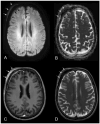
Figure 1.
Figure 1.
Extra- (white arrows A–D) and intracranial (black arrows A–D) transosseous lesion with diffusion restriction; particularly in the extracranial component, along with very low signal intensity on T2-weighted images and avid, nearly homogeneous contrast enhancement of the extra- and intracranial tumour components.
Figure 2.
Figure 2.
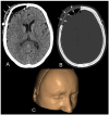
The preoperative CT scan revealed the extracranial subgaleal soft tissue component of the tumour (arrows A) and tiny calcifications along the thickened and enhancing right frontal dura (arrow B). However, the bone marrow of the frontal skull appeared unaffected.
Figure 3.
Figure 3.
First postoperative computed tomography scan after macroscopic complete resection of the intra- and extracranial tumour components, along with the infiltrated frontal bone, with normal postoperative findings: including intracranial extraaxial gas inclusions and a small extraaxial hygroma with residual lavage fluid (black arrows in A) and mild extracranial soft tissue swelling (arrows in A). B shows the patient-specific implant used to cover the frontal bone defect. Figure 3C shows the 3D-assisted cranioplasty with the skin covered.
Figure 4.
Figure 4.
Follow-up magnetic resonance imaging (MRI) 3 months after gross total resection. The brain MRI revealed a soft tissue mass at precisely the same subgaleal location as the primary tumour (white arrows A–E), with new invasion into the adjacent subcutaneous fat and skin. The recurrent tumour had expanded into adjacent anatomical regions, involving the right frontotemporal region, involving the parotid space, directly infiltrating both the superficial and deep lobes of the right parotid gland, and extending to the posterior right masticator space. The MRI characteristics were similar to those of the primary tumour, raising the possibility of intraoperative tumour seeding. The black arrows in A–E point to the patient’s specific implant with primary reactive granulation tissue in the course of the cranioplasty.
Figure 5.
Figure 5.
(A and B) Hematoxylin and eosin-stained slices show low- and high-power views of small- to medium-sized lymphoma cells with remnants of residual follicles. (C) The neoplastic B cells reveal an immunohistochemical expression of CD20 antibody. (D) Evaluation of Ki-67 shows a low proliferative activity displayed within the dense lymphoid infiltrates.

References

    1. Desjardins C, Larrieu-Ciron D, Choquet S, et al. Chemotherapy is an efficient treatment in primary CNS MALT lymphoma. J Neurooncol. 2022;159:151-161. 10.1007/s11060-022-04052-1 - DOI - PubMed
    1. Walewska R, Eyre TA, Barrington S, et al. ; BSH Committee. Guideline for the diagnosis and management of marginal zone lymphomas: a British Society of Haematology Guideline. Br J Haematol. 2024;204:86-107. 10.1111/bjh.19064 - DOI - PubMed
    1. Razaq W, Goel A, Amin A, Grossbard ML. Primary central nervous system mucosa-associated lymphoid tissue lymphoma: case report and literature review. Clin Lymphoma Myeloma. 2009;9:E5-E9. 10.3816/CLM.2009.n.052 - DOI - PubMed
    1. Flospergher E, Marino F, Calimeri T, et al. Primary central nervous system marginal zone lymphoma. Br J Haematol. 2024;204:31-44. 10.1111/bjh.19238 - DOI - PubMed
    1. Tu P h, Giannini C, Judkins AR, et al. Clinicopathologic and genetic profile of intracranial marginal zone lymphoma: a primary low-grade CNS lymphoma that mimics meningioma. J Clin Oncol. 2005;23:5718-5727. 10.1200/JCO.2005.17.624 - DOI - PubMed

Publication types

LinkOut - more resources