. 2025 Jul 23:43:102170.
doi: 10.1016/j.bbrep.2025.102170. eCollection 2025 Sep.

PMab-322: a novel anti-hippopotamus podoplanin monoclonal antibody for multiple applications

Affiliations

PMab-322: a novel anti-hippopotamus podoplanin monoclonal antibody for multiple applications

Haruto Yamamoto et al. Biochem Biophys Rep. .

Abstract

Podoplanin (PDPN) is a highly glycosylated type I transmembrane protein. PDPN expression is observed in various normal tissues, including lymphatic endothelial cells, kidney podocytes, and type I alveolar epithelial cells in the lungs. Monoclonal antibodies (mAbs) targeting PDPN across different animal species have facilitated the identification of PDPN-positive cells. To date, we have developed anti-PDPN mAbs for over 20 species. These antibodies suit various applications, including flow cytometry, immunoblotting, and immunohistochemistry. In this study, we generated an anti-hippopotamus PDPN (hipPDPN) mAb, PMab-322 (mouse IgG2a, kappa), using the Cell-Based Immunization and Screening (CBIS) method. PMab-322 exhibited strong reactivity to hipPDPN-overexpressed Chinese hamster ovary-K1 and demonstrated moderate affinity (K D: 4.4 × 10⁻⁸ M) in a flow cytometry-based measurement. PMab-322 specifically recognizes hipPDPN but does not cross-react with PDPN from 23 other species. Furthermore, PMab-322 successfully detected hipPDPN in both immunoblotting and immunohistochemistry. These findings highlight the potential of PMab-322 for pathological analyses of hippopotamus-derived tissues.

Keywords: CBIS method; Flow cytometry; Hippopotamus podoplanin; Immunoblotting; Immunohistochemistry; Monoclonal antibody.


Conflict of interest statement

The authors declare that they have no known competing financial interests or personal relationships that could have appeared to influence the work reported in this paper.

Figures

Fig. 1
Fig. 1
A schematic illustration of anti-hipPDPN mAbs production. (A) CHO/MAP16-hipPDPN was immunized into BALB/cAJcl mice. (B) The spleen cells were fused with P3U1 cells. (C) To select anti-hipPDPN mAb-producing hybridomas, the supernatants were screened by flow cytometry using CHO/PA16-hipPDPN and CHO–K1. (D) After limiting dilution, anti-hipPDPN mAbs were cloned by limiting dilution. PMab-322 (mouse IgG2a, kappa) was finally established.
Fig. 2
Fig. 2
Flow cytometric analysis of PMab-322 against CHO/PA16-hipPDPN. (A) CHO/PA16-hipPDPN and CHO–K1 were treated with 10–0.01 μg/mL of PMab-322 (red line) or blocking buffer (black line), followed by Alexa Fluor 488-conjugated anti-mouse IgG. (B) CHO/PA16-hipPDPN was suspended in 100 μL serially diluted PMab-322. Then, cells were treated with Alexa Fluor 488-conjugated anti-mouse IgG. Fluorescence data were subsequently collected using the SA3800 Cell Analyzer. The dissociation constant (KD) of PMab-322 was determined by GraphPad PRISM 6.
Fig. 3
Fig. 3
Specificity of PMab-322 against 24 species PDPN-overexpressed CHO–K1. (A) Twenty-four species PDPN-overexpressed CHO–K1 cell lines were treated with 10 μg/mL of PMab-322 (red line) or blocking buffer (black line), followed by Alexa Fluor 488-conjugated anti-mouse IgG. (B) The expression of each PDPN was confirmed by corresponding anti-PDPN mAbs (green line, 10 μg/mL). Then, Alexa Fluor 488-conjugated anti-mouse IgG or anti-rat IgG were treated. Fluorescence data were collected using the SA3800 Cell Analyzer. Note that the recognition of CHO/PA16-hipPDPN by NZ-1 was mediated by the reaction to PA16-tag.
Fig. 4
Fig. 4
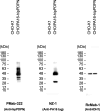
Detection of hipPDPN by immunoblotting. The membranes, on which cell lysates of CHO–K1 and CHO/PA16-hipPDPN were transferred, were incubated with 1 μg/mL of PMab-322, 1 μg/mL of NZ-1, or 1 μg/mL of RcMab-1. The membranes were incubated with horseradish peroxidase-conjugated anti-mouse (for PMab-322) or horseradish peroxidase-conjugated anti-rat immunoglobulins (for NZ-1 and RcMab-1). Chemiluminescence signals were developed and detected with a Sayaca-Imager.
Fig. 5
Fig. 5
Immunohistochemistry of paraffin-embedded sections of CHO/PA16-hipPDPN and CHO–K1. The sections of CHO/PA16-hipPDPN (A) and CHO–K1 (B) were treated with 0.1 μg/mL of PMab-322. The staining was carried out using BenchMark ULTRA PLUS with the ultraView Universal DAB Detection Kit. Scale bar = 100 μm.
Supplementary Fig. S1
The quantification of the PMab-322 reactivity in CHO–K1 cells, which overexpress PDPN derived from various species. The values (geometric mean of PMab-322 / geometric mean of each control mAb, Fig. 3) were determined.
figs2
figs2

Similar articles

References

    1. Kato Y., Fujita N., Kunita A., Sato S., Kaneko M., Osawa M., Tsuruo T. Molecular identification of Aggrus/T1alpha as a platelet aggregation-inducing factor expressed in colorectal tumors. J. Biol. Chem. 2003;278:51599–51605. doi: 10.1074/jbc.M309935200. - DOI - PubMed
    1. Suzuki H., Kaneko M.K., Kato Y. Roles of podoplanin in malignant progression of tumor. Cells. 2022;11 doi: 10.3390/cells11030575. - DOI - PMC - PubMed
    1. Hirakawa S., Hong Y.K., Harvey N., Schacht V., Matsuda K., Libermann T., Detmar M. Identification of vascular lineage-specific genes by transcriptional profiling of isolated blood vascular and lymphatic endothelial cells. Am. J. Pathol. 2003;162:575–586. doi: 10.1016/s0002-9440(10)63851-5. - DOI - PMC - PubMed
    1. Petrova T.V., Mäkinen T., Mäkelä T.P., Saarela J., Virtanen I., Ferrell R.E., Finegold D.N., Kerjaschki D., Ylä-Herttuala S., Alitalo K. Lymphatic endothelial reprogramming of vascular endothelial cells by the Prox-1 homeobox transcription factor. Embo j. 2002;21:4593–4599. doi: 10.1093/emboj/cdf470. - DOI - PMC - PubMed
    1. Breiteneder-Geleff S., Matsui K., Soleiman A., Meraner P., Poczewski H., Kalt R., Schaffner G., Kerjaschki D. Podoplanin, novel 43-kd membrane protein of glomerular epithelial cells, is down-regulated in puromycin nephrosis. Am. J. Pathol. 1997;151:1141–1152. - PMC - PubMed

LinkOut - more resources