. 2025 May 23;4(2):217-236.
doi: 10.1515/nipt-2025-0005. eCollection 2025 Jun.

Pannexin-1 channels, extracellular ATP, and purinergic receptors are essential for CCR5/CXCR4 clustering and HIV entry

Affiliations

Pannexin-1 channels, extracellular ATP, and purinergic receptors are essential for CCR5/CXCR4 clustering and HIV entry

David Ajasin et al. NeuroImmune Pharm Ther. .

Abstract

Objective: The Human Immunodeficiency Virus-1 (HIV) cell entry has been well characterized with the identification of CD4 as the main receptor and CXCR4 and CCR5 as co-receptors for the virus. However, how the virus uses the cell machinery for entry and infection is still a work-in-progress. Previously, we identified that the Pannexin-1 (Panx-1) channel, extracellular ATP, and purinergic receptors axis are essential for HIV entry and replication in macrophages, but the mechanisms were not fully explored.

Methods: Electrophysiology, ATP quantifications, confocal, HIV entry and replication experiments were used to determine the role of Panx-1 channels in HIV entry.

Results: Here, we identified that HIV or gp120 induces Panx-1 channel opening in association with ATP secretion, purinergic activation, and CCR5/CXCR4/actin clustering to enable HIV entry. Blocking Panx-1 channel opening, ATP secretion, or purinergic signaling prevented co-receptor clustering, HIV entry, and subsequent replication in multiple cell types.

Conclusion: We conclude that gp120 binding to the cell induces Panx-1 opening to promote the clustering of CCR5 or CXCR4 to the site of CD4-gp120 contact to aid viral entry.

Keywords: AIDS; HIV-1; cure; hemichannels; viral reservoirs.


Conflict of interest statement

Conflict of interest: The authors have no financial conflicts of interest.

Figures
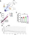
Figure 1:
Figure 1:
gp120 protein induces opening of Panx-1 channels, (A) schematic of electrophysiological recording from U87-CD4-CCR5 cells. (B) Current/voltage (I/V) curves from U87CD4CCR5 cell patch clamp experiments in control (CTRL), ATP, gp120 only, and gp120+ATP, n=3. (C) Quantification of fold change relative to peak conductance (***p≤0.005 compared to control conditions, n=8). (D) Representative recording of patch-clamped U87CD4CCR5 cells treated with gp120+1 µM ATP over 4,000 s. Statistics: nonparametric t-test, n=8, ***p value ≤0.001.
Figure 2:
Figure 2:
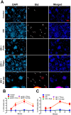
Exposure of PBMCs to gp120 or HIV induces Panx-1 channel opening. (A) Representative image of PHA-activated PBMCs plated and subjected to the following pre-treatment for 30 min (PBS-only for control and gp120/HIV only), 500 µM Probenecid, 300 µM Panx-1 mimetic peptide, 10Panx peptide, and 300 µM scrambled peptide (Scr pep). Afterwards, cells were exposed to gp120 (200 ng/mL) or NL4-3 (10 ng/mL). Etd was added, and Etd uptake was monitored with live cell imaging. Image is at 10 min post-addition of Etd, n=3. White arrows indicate cells with Etd uptake. (B) Quantification of the percentage of Etd uptake in all gp120-treated PBMCs relative to DAPI. (C) Quantification of percentage of Etd uptake in all HIV infected PBMCs relative to DAPI displayed as % of Etd uptake cells is determined by number of cells positive for Etd/total number of cells (DAPI positive) × 100. Statistics: area under the curve p value ≤0.05 from 10 to 30 min; control versus. gp120/HIV, control versus. gp120/HIV+Scr pep, gp120/HIV versus. gp120/HIV+Prob/10Panx. Data are represented as mean±SEM, n=3.
Figure 3:
Figure 3:
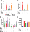
Blocking the Panx-1 channel inhibits ATP release in PBMCs, MDMs, and CEM-GFP cells. (A) PHA-activated PBMCs were subjected to the following pre-treatments for 30 min PBS-only (for control and HIV only), 500 µM Probenecid, 300 µM Panx-1 mimetic peptide, 10Panx peptide, and 300 µM scrambled peptide (Scr pep). Followed by replication of HIV after inoculation with NL4-3 (10 ng/mL), and media collected every 3 days until day 15 post-infection and ATP levels determined with ATP luciferase/luciferin-based enzyme assay, n=3. (B) MDMs were subjected to the following pre-treatments for 30 min PBS-only (for control and HIV only), 500 µM Probenecid, 300 µM Panx-1 mimetic peptide, 10-Panx peptide, and 300 µM scrambled peptide (Scr pep). Followed by replication of HIV after inoculation with pNL(AD8) (10 ng/mL) and media collected on days 3, 7, 11, 15, 19, 23, and 28 post-infection, followed by ATP luciferase/luciferin-based enzyme assay to determine ATP levels, n=3. (C) CEM-GFP cells were subjected to the following pre-treatments for 30 min media-only (for control and HIV only), 500 µM Probenecid (Prob), 300 µM Panx-1 mimetic peptide, (10Panx), and 300 µM scrambled peptide (Scr pep). Followed by replication of HIV after inoculation with NL4-3 (10 ng/mL) for 15 days, and media collected every 3 days until day 15 post-infection. Extracellular ATP levels in media were determined with ATP quantification assay using luciferase/luciferin-based enzyme assay, n=3. Statistics: area under the curve show significant difference p value <0.05 control versus. HIV, and control versus. HIV+Scr pep, also, there is significant difference, p value <0.05 HIV versus. HIV+Prob and HIV versus. HIV+ 10Panx-1. Data are represented as mean±SD.
Figure 4:
Figure 4:
Pannexin-1 and released extracellular ATP contribute to HIV entry in MDMs and cell lines. (A) Pre-treated MDMs were infected with β-lactamase containing pseudo-typed virus (pWT/BaL-pMM310) for HIV entry assay, n=12. (B) Pre-treated THP-1 cells were infected with β-lactamase containing pseudo-typed virus (pWT/BaL-pMM310) for HIV entry assay, n=4. (C) MDMs from an individual was pre-treated with BzATP (300 µM), ATPγS (15 µM), oATP (200 µM), 10-Panx peptide (300 µM), and scrambled (Scr) peptide (300 µM), and inoculated with β-lactamase containing pseudo-typed virus (pWT/BaL-pMM310, 200 ng/mL) to test for HIV entry at 4-, 8-, and 12-h post-inoculation, n=4. % of HIV-1 Entry is determined as; number of cells positive at 460 nm/number of cells positive at 528 nm × 100. (D) Relative Light Unit (RLU) of pre-treated TZMbL cells inoculated with 10 ng/mL pNL(AD8), to HIV entry on viral transcription, n=3. Statistics: nonparametric t-test for figures A, B, D, and Two-Way ANOVA for figure C. Data are represented as mean±SD, *p≤0.005 compared to controls and #p≤0.005 compared to HIV.
Figure 5:
Figure 5:
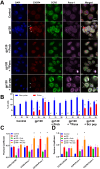
gp120 induce CXCR4 clustering in PBMCs through Panx-1 opening. (A) Confocal images of PHA-activated and pre-treated PBMCs exposed to LAV gp120 (200 ng/mL) and stained for DAPI, CXCR4, CCR5, and Panx-1 proteins to visualize co-receptor distribution in all conditions examined. Arrows indicate co-receptor clustering, n=3, Scale bar=20 µm. (B) Quantification of cells with CXCR4 clustering in response to examined conditions from confocal imaging in (A). Statistics: Two-Way ANOVA, data are represented as mean±SD, *p≤0.005 compared to untreated and #p≤0.005 compared to gp120 only condition. (C) Pearson colocalization coefficient of gp120-LAV treated cells in (A) above shows Panx-1 colocalizing (>0.5) in gp120 only and gp120+Scr peptide conditions and no colocalization in Probenecid and 10Panx peptide conditions. (D) Pearson colocalization coefficient of BaL gp120 treated cells in Supplementary Figure 5 shows Panx-1 colocalizing (>0.5) in gp120 only and gp120+Scr peptide conditions and no colocalization in Probenecid and 10Panx peptide conditions.
Figure 6:
Figure 6:
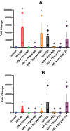
Reverse transcriptase (RT) product synthesis is reduced by blocking of the Panx-1 channel opening. Fold change determined via qPCR ascertains the level of early (A) or late (B) RT products synthesized from pre-treated and HIV-infected macrophages at 48- and 72-h post-infection, n=3. Data are represented as mean±SD, *p≤0.005 compared to control.
Figure 7:
Figure 7:
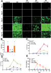
Blocking the Panx-1 channel prevents HIV replication in CEM-GFP, PBMCs, and MDMs. (A) Epifluorescence imaging of multi-day replication (15 days) of pre-treated and NL4-3 infected CEM-GFP cells, with GFP expression indicative of HIV replication. Images collected every 3 days, n=3, Scale bar=100 µm. (B) Quantification of GFP signal from all images from the experiment shown in (A) above using plate reader, n=3 and (A.U) = arbitrary unit. (C–D) p24 ELISA from pre-treated and HIV-infected CEM-GFP cells (C) and PBMCs (D) over 15 days of replication. (E) p24 ELISA from pre-treated and HIV-infected MDMs over 28 days of replication. Data are represented as mean±SD, n=3.

Similar articles

References

    1. Osega CE, Bustos FJ, Arriagada G. From entry to the nucleus: how retroviruses commute. Annu Rev Virol. 2024;11:89–104. doi: 10.1146/annurev-virology-100422-023502. - DOI - PubMed
    1. Kwong PD, Wyatt R, Robinson J, Sweet RW, Sodroski J, Hendrickson WA. Structure of an HIV gp120 envelope glycoprotein in complex with the CD4 receptor and a neutralizing human antibody. Nature. 1998;393:648–59. doi: 10.1038/31405. - DOI - PMC - PubMed
    1. Orloff GM, Orloff SL, Kennedy MS, Maddon PJ, McDougal JS. Penetration of CD4 T cells by HIV-1. The CD4 receptor does not internalize with HIV, and CD4-related signal transduction events are not required for entry. J Immunol. 1991;146:2578–87. doi: 10.4049/jimmunol.146.8.2578. - DOI - PubMed
    1. Burdick RC, Li C, Munshi M, Rawson JMO, Nagashima K, Hu WS, et al. HIV-1 uncoats in the nucleus near sites of integration. Proc Natl Acad Sci U S A. 2020;117:5486–93. doi: 10.1073/pnas.1920631117. - DOI - PMC - PubMed
    1. Vink C, van Gent DC, Plasterk RH. Integration of human immunodeficiency virus types 1 and 2 DNA in vitro by cytoplasmic extracts of Moloney murine leukemia virus-infected mouse NIH 3T3 cells. J Virol. 1990;64:5219–22. doi: 10.1128/jvi.64.10.5219-5222.1990. - DOI - PMC - PubMed

LinkOut - more resources