. 2025 Jul 27;17(7):105136.
doi: 10.4240/wjgs.v17.i7.105136.

Comparison of endoscopic ultrasonography features and pathological staging of gastric inflammatory fibroid polyps

Affiliations

Comparison of endoscopic ultrasonography features and pathological staging of gastric inflammatory fibroid polyps

Fen-Ming Zhang et al. World J Gastrointest Surg. .

Abstract

Background: The diagnosis of gastric inflammatory fibroid polyps (IFPs) mainly depends on pathological confirmation after endoscopic or surgical treatment. Gastric IFP have typical manifestations under endoscopic ultrasonography (EUS), but atypical EUS features have also been reported. Previous studies have found that atypical features of gastric IFPs observed under EUS have corresponding histological manifestations. At present, there is no study elaborating the EUS manifestations of gastric IFPs at different pathological stages. We hypothesize that gastric IFPs at different pathological stages may have different EUS features.

Aim: To describe EUS features of gastric IFPs and compare with their pathological characteristics.

Methods: Clinical data of 53 inpatients with pathologically diagnosed gastric IFPs after endoscopic treatment were collected. All patients underwent preoperative EUS. We analyzed the EUS characteristics of the lesions and compared with the pathological characteristics and staging of the resected specimens.

Results: Most gastric IFPs showed medium-low echo (67.9%), homogeneous echo (90.6%), and unclear boundaries (83%), and involved the second and third layers of the gastric wall (69.8%) under EUS. The echogenicity level and echo homogeneity were significantly correlated with the pathological stage of gastric IFP. Gastric IFPs in the nodular stage presented hypoechoic and homogeneous echo. Gastric IFPs in the fibrovascular stage mostly showed medium-low echo and homogeneous echo. Gastric IFPs in the sclerotic stage showed different echogenicity levels and echo homogeneity. The accuracy of EUS in diagnosing gastric IFPs was 66.0% (35/53), and the accuracy in determining the origin layer of gastric IFPs was 73.4% (39/53).

Conclusion: Gastric IFPs at different pathological stages have different EUS features. In order to improve the diagnostic rate, it is necessary to combine EUS with EUS-guided fine-needle aspiration or artificial intelligence.

Keywords: Endoscopic ultrasonography; Fibrovascular stage; Gastric inflammatory fibroid polyps; Nodular stage; Sclerotic stage.


Conflict of interest statement

Conflict-of-interest statement: The authors declare that they have no conflict of interest.

Figures

Figure 1
Figure 1
Different manifestations of gastric inflammatory fibroid polyps under endoscopic ultrasonography. A: The lesion originated from the third layer, with low and homogeneous echo and indistinct margin, and diagnosed as a gastric neuroendocrine tumor by preoperative endoscopic ultrasonography (EUS), while the pathological results suggested a gastric inflammatory fibroid polyps (IFP) in the fibrovascular stage; B: The lesion originated from the second layer, with medium-low and homogeneous echo and distinct margin, and diagnosed as gastric polyp preoperatively by EUS while the pathological results suggested a gastric IFP in the fibrovascular stage; C: The lesion originated from the second layer, with medium-high and homogeneous echo and indistinct margin, and diagnosed as a gastric adenoma by preoperative EUS, while the pathological results suggested a gastric IFP in the fibrovascular stage; D: The lesion involved both the second and third layers, with high and heterogeneous echo and indistinct margin, and was diagnosed as a ectopic pancreas of the stomach by EUS preoperatively, while the pathological results suggested a gastric IFP in the fibrovascular stage.
Figure 2
Figure 2
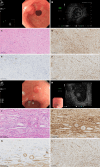
Endoscopic, ultrasonic endoscopic and pathological characteristics of gastric inflammatory fibroid polyps. A-F: Nodular stage; A: General endoscopic features: A submucosal protrusion on the anterior wall of the gastric antrum near the pylorus, with smooth and intact surface mucosa; B: Endoscopic ultrasonography (EUS) features: The lesion originated from the third layer, with low echo, homogeneous echo and indistinct margin; C: Pathological characteristics: Immature fibroblastic-like spindle cell proliferation, few inflammatory cells such as eosinophils, and a small amount of thin-walled vascular proliferation, hematoxylin-eosin (HE): 200 ×; D: Immunohistochemical cluster of differentiation (CD) 34 +: 200 ×; E: Immunohistochemical smooth muscle actin (SMA) +: 200 ×; F: Immunohistochemical vimentin +: 200 ×; G-L: Fibrovascular stage; G: General endoscopic features: A submucosal protrusion on the lesser curvature of the gastric antrum, with hyperemia of the surface mucosa; H: EUS features: Lesion originated from the second layer, with medium-low echo, homogeneous echo and indistinct margin; I: Pathological characteristics: Mature fibroblastic-like spindle cell proliferation, thin- and thick-walled blood vessels are visible, and there is a significant eosinophil proliferation around the blood vessels. The proliferating spindle cells exhibit a classic “onion skin” pattern around small blood vessels, HE: 200 ×; J: Immunohistochemical CD34 +: 200 ×; K: Immunohistochemical SMA +: 200 ×; L: Immunohistochemical vimentin + 200 ×.
Figure 3
Figure 3
Endoscopic, ultrasonic endoscopic and pathological characteristics of sclerotic stage gastric inflammatory fibroid polyps. A: General endoscopic features: A submucosal protrusion on the anterior wall of the gastric antrum, the surface mucosa was congested with ulcer formation on the top; B: Endoscopic ultrasonography features: The lesion involved both the second and third layer, with medium-high echo, heterogeneous echo and indistinct margin; C: Pathological features: Collagen fiber proliferation with concomitant hyalinization, thickened blood vessels, and extensive eosinophil infiltration, hematoxylin-eosin: 200 ×; D: Immunohistochemical cluster of differentiation 34 +: 200 ×; E: Immunohistochemical smooth muscle actin + 200 ×.

Similar articles

References

    1. Kim YI, Kim WH. Inflammatory fibroid polyps of gastrointestinal tract. Evolution of histologic patterns. Am J Clin Pathol. 1988;89:721–727. - PubMed
    1. Matsushita M, Hajiro K, Okazaki K, Takakuwa H. Gastric inflammatory fibroid polyps: endoscopic ultrasonographic analysis in comparison with the histology. Gastrointest Endosc. 1997;46:53–57. - PubMed
    1. Inayat F, Ur Rahman A, Wahab A, Riaz A, Zahid E, Bejarano P, Pimentel R. Gastric Inflammatory Fibroid Polyp: A Rare Cause of Occult Upper Gastrointestinal Bleeding. J Investig Med High Impact Case Rep. 2020;8:2324709620936840. - PMC - PubMed
    1. Nagao S, Tsuji Y, Sakaguchi Y, Ushiku T, Koike K. Inflammatory fibroid polyp mimicking an early gastric cancer. Gastrointest Endosc. 2020;92:217–218. - PubMed
    1. Garmpis N, Damaskos C, Garmpi A, Georgakopoulou VE, Sakellariou S, Liakea A, Schizas D, Diamantis E, Farmaki P, Voutyritsa E, Syllaios A, Patsouras A, Sypsa G, Agorogianni A, Stelianidi A, Antoniou EA, Kontzoglou K, Trakas N, Dimitroulis D. Inflammatory Fibroid Polyp of the Gastrointestinal Tract: A Systematic Review for a Benign Tumor. In Vivo. 2021;35:81–93. - PMC - PubMed

LinkOut - more resources