Case Reports
. 2025 Jul 27;17(7):104985.
doi: 10.4240/wjgs.v17.i7.104985.

Multiple jejunal diverticulosis, from an anatomical and histological view: A case report

Affiliations
Case Reports

Multiple jejunal diverticulosis, from an anatomical and histological view: A case report

Patricia Schmidt et al. World J Gastrointest Surg. .

Abstract

Background: Here, we report a case of jejunal diverticulosis from an anatomical and histological view. During the "Gross Anatomy course," we found multiple jejunal diverticula along a total length of 208 cm of intestine.

Case summary: After opening the intestinal tract, we counted 232 jejunal diverticulum entry points with a diameter of up to 2 cm and observed connections between the diverticula that created shortcuts between two distinct intestinal parts. Interestingly, we observed an extreme longitudinal striation on the intestinal parts hosting diverticula. Thorough vessel preparation utilizing a dissecting microscope confirmed that all investigated arteriae rectae ended in a diverticulum. Histological and immunohistochemical investigations revealed that intestinal villi of diverticula were smaller and less prominent than control tissue and that the stratum longitudinale, as well as the stratum circular, were much thinner in the diverticula compared to control tissue. Neither submucosal nor mesenteric plexus could be detected in the diverticula. However, vasoactive intestinal peptide-positive nerve fibers and villin-positive brush border could only be detected in control tissue. This indicates that jejunal diverticulosis is associated with abnormalities of the smooth muscles and a disorder of innervation.

Conclusion: Jejunal diverticulosis originates from mesenteric vessels, featuring smooth muscle changes, absent innervation, and thinning of tissue layers.

Keywords: Case report; Diverticulosis; Intestinal abnormalities; Jejunal diverticula; Translational medicine.


Conflict of interest statement

Conflict-of-interest statement: The authors have no conflicts of interest to declare.

Figures
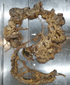
Figure 1
Figure 1
Overview of intestine harboring diverticula. Intestinal tract including jejunum, ileum, and colon. Jejunum hosting diverticula over a length of 208 cm. Arrows mark superior mesenteric artery. Asterisks mark cut connection of artery branch due to preparation and visualization. Arrowheads mark inferior mesenteric artery.
Figure 2
Figure 2
Diverticula. A-C: Representative diverticula; D: Shortcut between two intestinal parts provided by diverticula.
Figure 3
Figure 3
Longitudinal striation of jejunum with diverticula. A: Intestinal part hosting diverticula showed remarkable longitudinal striation. Black box marks magnified part shown in B and C; B and C: Magnification of longitudinal striation; D: Intestinal part without diverticula.
Figure 4
Figure 4
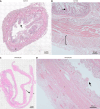
Microscopic investigation of longitudinal striation of jejunum with diverticula. A and B: Hematoxylin and eosin (H&E)-stained diverticula and control tissue; C and D: Magnification of H&E-stained diverticula and control tissue. Arrows mark indentations responsible for longitudinal striation.
Figure 5
Figure 5
Lumen of intestine and diverticula. A: Intestinal tract was cut longitudinally. Arrows mark diverticula entry points; B: Representative diverticula were opened; C and D: Diverticula entry points pictured from intestinal site (C) and from diverticulum site (D); E: Large diverticulum entry points with a diameter of 2 cm.
Figure 6
Figure 6
Relationship between vessels and diverticula. Representative part of the jejunum (8 cm) with diverticula was cut out and dissected utilizing a dissecting microscope. All remaining fat tissue and intestine covering tunica serosa was removed, vessels were stretched out, and entry sites were exposed. Arrows mark arteria recta entering diverticula (marked with an asterisk). All investigated vessels end in diverticula, and all investigated diverticula were supplied by a vessel.
Figure 7
Figure 7
Histological investigation of diverticula. A and C: Hematoxylin and eosin (H&E)-stained control tissue (A) and diverticula (C). Arrows mark intestinal villi. Asterisks mark stratum longitudinale; B and D: H&E-stained control tissue (B) and diverticula (D) in a higher magnification. 1: Stratum circular; 2: Stratum longitudinale. Arrows mark intestinal villi.
Figure 8
Figure 8
Immunohistochemical investigation of diverticula. A and B: Immunohistochemical staining with smooth muscle actin antibody of control tissue (A) and diverticula (B); C-F: Investigation of intestinal villi in control tissue (C and E) and diverticula (D and F) utilizing vasoactive intestinal peptide (VIP; C and D) and villin (E and F) to mark VIP-positive fibers and brush border, respectively. 1: Intestinal villi; 2: Lamina muscularis mucosa; 3: Lamina submucosae; 4: Stratum circular; 5: Stratum longitudinale; 6: Adventitia/serosa.
Figure 9
Figure 9
Immunohistochemical investigation of submucosal plexus in diverticula. A-H: Detection of submucosal plexus in control tissue (A, C, E, and G) and diverticula (B, D, F, and H) utilizing choline acetyltransferase (ChAT) (A and B), protein gene product 9.5 (PGP9.5) (C and D), substance P (SP) (E and F) and neuronal nitric oxide synthase (nNOS) (G and H) antibody. Arrows mark positive cells in the plexus.
Figure 10
Figure 10
Immunohistochemical investigation of myenteric plexus in diverticula. A-H: Detection of myenteric plexus in control tissue (A, C, E, and G) and diverticula (B, D, F, and H) utilizing choline acetyltransferase (ChAT) (A and B), protein gene product 9.5 (PGP9.5) (C and D), substance P (SP) (E and F) and neuronal nitric oxide synthase (nNOS) (G and H) antibody. Arrows mark positive cells in the plexus.

Similar articles

References

    1. Parks TG. Natural history of diverticular disease of the colon. Clin Gastroenterol. 1975;4:53–69. - PubMed
    1. Painter NS, Burkitt DP. Diverticular disease of the colon: a deficiency disease of Western civilization. Br Med J. 1971;2:450–454. - PMC - PubMed
    1. Hobson KG, Roberts PL. Etiology and pathophysiology of diverticular disease. Clin Colon Rectal Surg. 2004;17:147–153. - PMC - PubMed
    1. Comparato G, Pilotto A, Franzè A, Franceschi M, Di Mario F. Diverticular disease in the elderly. Dig Dis. 2007;25:151–159. - PubMed
    1. Wedel T, Barrenschee M, Lange C, Cossais F, Böttner M. Morphologic Basis for Developing Diverticular Disease, Diverticulitis, and Diverticular Bleeding. Viszeralmedizin. 2015;31:76–82. - PMC - PubMed

Publication types

LinkOut - more resources