Review
. 2025 Jul 23;17(7):e88584.
doi: 10.7759/cureus.88584. eCollection 2025 Jul.

Practical Applications of Lung and Diaphragm Ultrasound in the Intensive Care Unit: An Updated Narrative Review

Affiliations
Review

Practical Applications of Lung and Diaphragm Ultrasound in the Intensive Care Unit: An Updated Narrative Review

Keith Killu et al. Cureus. .

Abstract

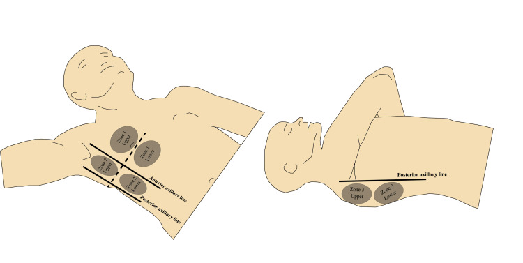
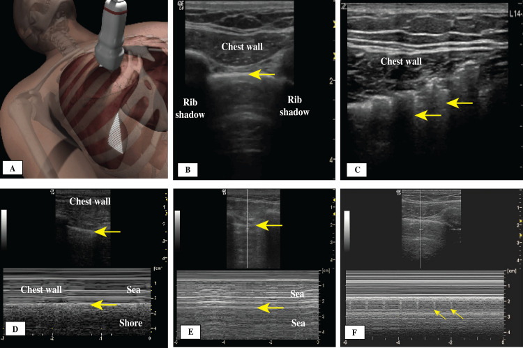
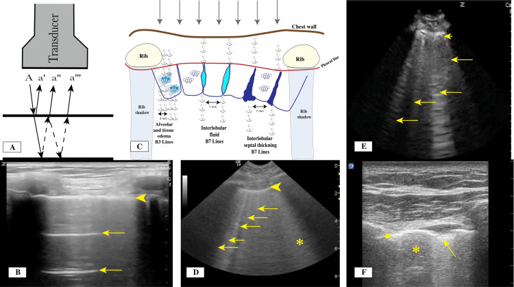
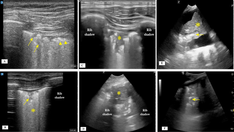
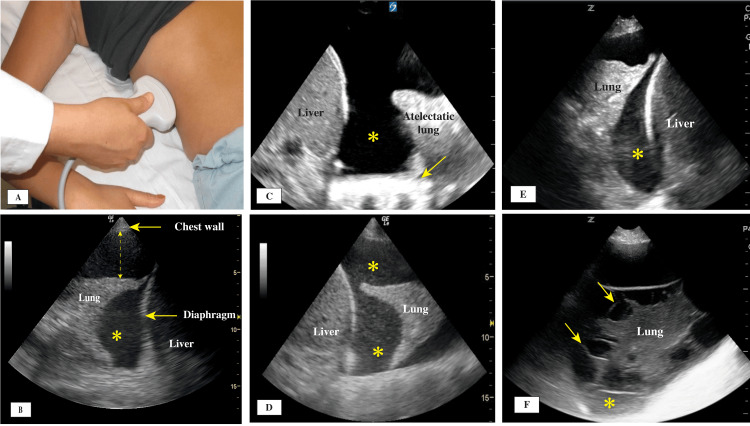
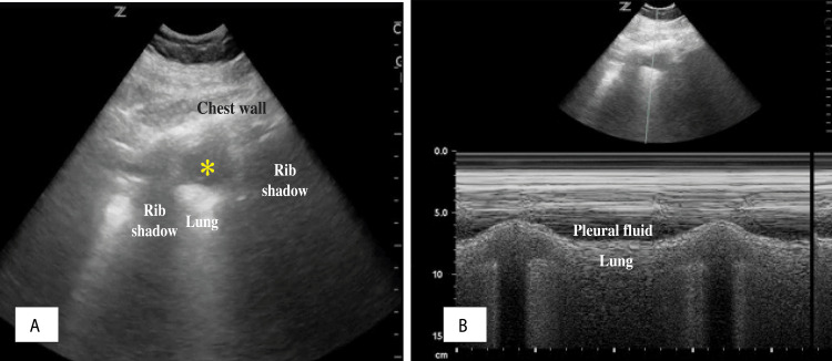
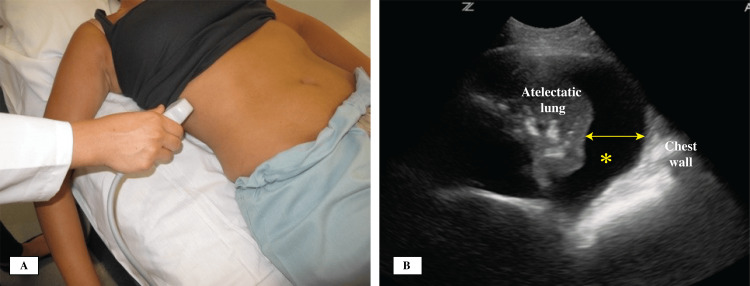
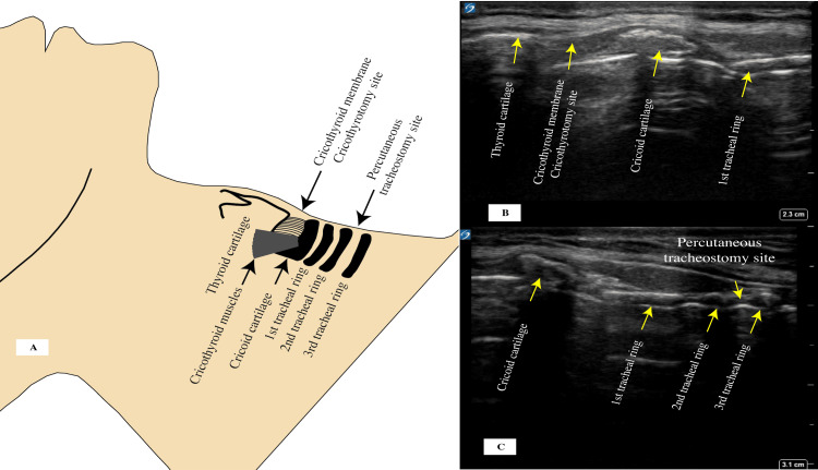
Lung ultrasound (LUS) has evolved significantly during the past few decades. Its use has been integrated into daily practices of intensive care units (ICUs) worldwide, and it has proven to be a valuable tool in the assessment and management of patients with respiratory failure caused by lung, pleural, diaphragmatic, and other diseases. LUS techniques are becoming increasingly standardized, which can help in interpreting data and improving patients' outcomes. In this narrative review, the focus was on the practical daily applications of lung, pleural, and diaphragmatic ultrasound with emphasis on different signs and artifacts that guide the interpretation of data and identification of disease. Discussions and analysis from the new international guidelines were added to help close the gap in the use of LUS. This review is intended to serve as a practical guide for using bedside ultrasound in evaluating patients with shortness of breath and respiratory failure and to provide guidance to help providers manage patients and generate standardized reports. We start with an analysis of best practices and guidelines on performing an LUS exam in the ICU setting. This analysis is followed by data interpretation of findings starting at the pleural line and traveling deeper into the lung tissue. The review includes discussions of the diaphragm evaluation and its function and abnormalities, as well as common LUS-related procedures in the ICU, such as thoracentesis, tracheostomy, and cricothyrotomy.

Keywords: a-lines; acute respiratory distress syndrome (ards); b-lines; diaphragm ultrasound (d-usg); lung ultrasound (lus); percutaneous tracheostomy; pleural fluid (pf); pleural ultrasound; pneumonia; point-of-care ultrasound (pocus).


Conflict of interest statement

Conflicts of interest: In compliance with the ICMJE uniform disclosure form, all authors declare the following: Payment/services info: All authors have declared that no financial support was received from any organization for the submitted work. Financial relationships: All authors have declared that they have no financial relationships at present or within the previous three years with any organizations that might have an interest in the submitted work. Other relationships: All authors have declared that there are no other relationships or activities that could appear to have influenced the submitted work.

Figures

Figure 1
Figure 1. Lung zones.
Zone 1: upper and lower anterior; Zone 2: upper and lower lateral; Zone 3: upper and lower posterior. Credit: Image created by the authors.
Figure 2
Figure 2. (A) Initial transducer placement location; (B) Bat sign: normal thin pleural line (arrow) between two rib shadows; (C) E lines (arrows); (D) M-mode, seashore sign, pleural line (arrows); (E) M-mode, stratosphere sign, pleural line (arrows); (F) Lung pulse sign showing transmitted heart beats to the pleural line.
Figure 3
Figure 3. (A) Reverberation artifact mechanism for the horizontal A lines. (B) With the use of a linear transducer, a thin pleural line (arrowhead) and horizontal lines (A lines; arrows) are visible. (C) Proposed mechanism for the reverberation and formation of vertical lines (B lines). (D) With the use of a curvilinear transducer, a thin pleural line (arrowhead) with multiple vertical lines (B3 Lines) with 3 mm between visible lines can be seen in a patient with pulmonary edema. The asterisk indicates white lung with coalesced B Lines. (E) With the use of a curvilinear transducer, a thick pleural line (arrowhead) with multiple vertical lines (B7 lines) (arrows) with wider spaces between the B lines can be seen in a patient with thickening interlobular septa. (F) With the use of a linear transducer, an interrupted thin pleural line (arrowhead), subpleural consolidation (arrow), and skip area (asterisk) are visible.
Credit: Images A and C created by the authors.
Figure 4
Figure 4. (A) With use of a linear transducer, a thick pleural line (arrowheads) with multiple areas of subpleural consolidations (arrows) could be visualized. (B) A linear transducer also enabled visualizing a thick interrupted pleural line with subpleural consolidation (arrow) and numerous vertical lines forming the white lung sign (asterisk). (C) A linear transducer revealed a shred sign (asterisk) with interruptions and irregularity of the pleural line. (D) With use of a curvilinear transducer, a tissue-like pattern representing trans-lobar consolidation (asterisk) from pneumonia was shown. (E) A curvilinear transducer was used and showed dynamic air bronchogram with longitudinal bright shape (arrow), moving through the airway during the respiratory cycle, and collapsed lung tissue (asterisk). (F) A curvilinear transducer was used and revealed static air bronchogram, which usually resembles small circles that do not move with respiration.
Figure 5
Figure 5. (A) Posterolateral point area for transducer placement. (B) Pleural effusion with jellyfish sign. The dashed arrow measures the distance between the lung and chest wall for estimation of the pleural effusion size. (C) Simple anechoic pleural fluid (asterisk) and the thoracic spine sign (arrow). (D) Complex echoic pleural fluid with floating debris (plankton sign) (asterisks). (E) Complex echoic pleural fluid with debris accumulating in the dependent area owing to hemothorax (hematocrit sign; asterisks). (F) Complex echoic pleural fluid with septations (arrow) and thick debris accumulating in the dependent area (empyema; asterisk).
Figure 6
Figure 6. (A) Quad sign formed between the chest wall superiorly, pleural line inferiorly, and rib shadows on sides (asterisk); (B) M-mode of the quad sign showing the sinusoid sign.
Figure 7
Figure 7. (A) Illustration of the zone of apposition with the peritoneal and pleural surfaces. (B) Zone of apposition showing the pleural line (asterisk) reaching the diaphragm. (C) Diaphragm excursion measurement transducer placement. (D) M-mode showing the amplitude of the diaphragmatic excursion. (E) Transducer position and orientation to measure the diaphragmatic thickness. (F) M-mode of the diaphragm thickness changes with the respiratory cycle (yellow bands).
Credit: Image A created by the authors.
Figure 8
Figure 8. (A) Transducer placement in the posterior lateral zone to estimate pleural effusion size; (B) Pleural fluid size measurement with the widest diameter. Clear pleural fluid is marked with an asterisk.
Figure 9
Figure 9. (A) Cricothyroid membrane and anatomy for cricothyrotomy and percutaneous tracheostomy. (B) Ultrasound image showing the relation of the cricothyroid membrane to the thyroid and cricothyroid cartilages with the first and second tracheal rings. (C) Cricothyroid cartilage with the sub cricoid space and the first three tracheal rings.
Credit: Image A created by the authors.

Similar articles

  • [Volume and health outcomes: evidence from systematic reviews and from evaluation of Italian hospital data].
    Amato L, Colais P, Davoli M, Ferroni E, Fusco D, Minozzi S, Moirano F, Sciattella P, Vecchi S, Ventura M, Perucci CA. Amato L, et al. Epidemiol Prev. 2013 Mar-Jun;37(2-3 Suppl 2):1-100. Epidemiol Prev. 2013. PMID: 23851286 Italian.
  • Ventilator Management(Archived).
    Mora Carpio AL, Mora JI. Mora Carpio AL, et al. 2023 Mar 27. In: StatPearls [Internet]. Treasure Island (FL): StatPearls Publishing; 2025 Jan–. 2023 Mar 27. In: StatPearls [Internet]. Treasure Island (FL): StatPearls Publishing; 2025 Jan–. PMID: 28846232 Free Books & Documents.
  • Systemic Inflammatory Response Syndrome.
    Baddam S, Burns B. Baddam S, et al. 2025 Jun 20. In: StatPearls [Internet]. Treasure Island (FL): StatPearls Publishing; 2025 Jan–. 2025 Jun 20. In: StatPearls [Internet]. Treasure Island (FL): StatPearls Publishing; 2025 Jan–. PMID: 31613449 Free Books & Documents.
  • Health professionals' experience of teamwork education in acute hospital settings: a systematic review of qualitative literature.
    Eddy K, Jordan Z, Stephenson M. Eddy K, et al. JBI Database System Rev Implement Rep. 2016 Apr;14(4):96-137. doi: 10.11124/JBISRIR-2016-1843. JBI Database System Rev Implement Rep. 2016. PMID: 27532314
  • Management of urinary stones by experts in stone disease (ESD 2025).
    Papatsoris A, Geavlete B, Radavoi GD, Alameedee M, Almusafer M, Ather MH, Budia A, Cumpanas AA, Kiremi MC, Dellis A, Elhowairis M, Galán-Llopis JA, Geavlete P, Guimerà Garcia J, Isern B, Jinga V, Lopez JM, Mainez JA, Mitsogiannis I, Mora Christian J, Moussa M, Multescu R, Oguz Acar Y, Petkova K, Piñero A, Popov E, Ramos Cebrian M, Rascu S, Siener R, Sountoulides P, Stamatelou K, Syed J, Trinchieri A. Papatsoris A, et al. Arch Ital Urol Androl. 2025 Jun 30;97(2):14085. doi: 10.4081/aiua.2025.14085. Epub 2025 Jun 30. Arch Ital Urol Androl. 2025. PMID: 40583613 Review.

References

    1. Ultrasound for routine clinical investigations. Pell RL. Ultrasonics. 1964;2:87–89.
    1. Relevance of lung ultrasound in the diagnosis of acute respiratory failure: the BLUE protocol. Lichtenstein DA, Mezière GA. Chest. 2008;134:117–125. - PMC - PubMed
    1. International evidence-based recommendations for point-of-care lung ultrasound. Volpicelli G, Elbarbary M, Blaivas M, et al. Intensive Care Med. 2012;38:577–591. - PubMed
    1. New international guidelines and consensus on the use of lung ultrasound. Demi L, Wolfram F, Klersy C, et al. J Ultrasound Med. 2023;42:309–344. - PMC - PubMed
    1. Proposal for international standardization of the use of lung ultrasound for patients with COVID- 19: a simple, quantitative, reproducible method. Soldati G, Smargiassi A, Inchingolo R, et al. J Ultrasound Med. 2020;39:1413–1419. - PMC - PubMed

LinkOut - more resources