

doi: 10.1002/ccr3.70682.
eCollection 2025 Aug.
Neonatal Epidermolytic Ichthyosis Caused by a KRT10 Mutation (c.467G>A, p.Arg156His): A Case Report
Affiliations
- PMID: 40741111
- PMCID: PMC12307240
- DOI: 10.1002/ccr3.70682
Item in Clipboard
Neonatal Epidermolytic Ichthyosis Caused by a KRT10 Mutation (c.467G>A, p.Arg156His): A Case Report
Clin Case Rep.
.
Abstract
We present a neonatal case of skin blisters and erythema. While epidermolysis bullosa was initially suspected, immunofluorescence antigen mapping and genetic testing confirmed epidermolytic ichthyosis, with a heterozygous pathogenic variant in the KRT10 gene (c.467G>A, p.Arg156His). A multidisciplinary approach is essential for accurate diagnosis and treatment of neonatal blistering conditions.
Keywords: blistering skin disease; congenital erythroderma; epidermolysis bullosa; epidermolytic hyperkeratosis; epidermolytic ichthyosis.
© 2025 The Author(s). Clinical Case Reports published by John Wiley & Sons Ltd.
Conflict of interest statement
The authors declare no conflicts of interest.
Figures

Clinical features in the affected newborn: (A) The patient's right upper limb showing diffuse erythroderma and areas of denuded skin (arrow), and (B) superficial blister formation (arrows) on the flexural surface of the lower limb.

Clinical features showing the characteristic thickening, scaling, and yellowish appearance of the skin (arrows): (A) on the hand, and (B) on the flexural surfaces of the lower extremities, most notably in the popliteal folds and around the ankles.
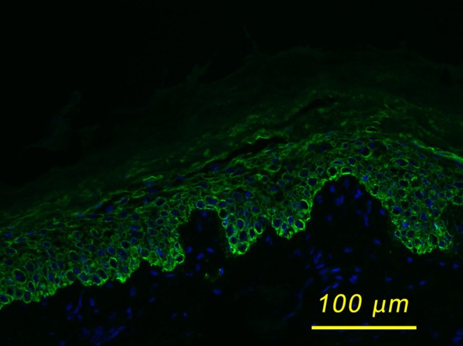
Immunofluorescence staining of the skin biopsy specimen obtained from a clinically affected area (lower left limb), using the LL001 antibody against KRT14, which highlights the basal keratinocytes. A distinct subcorneal cleft is observed, consistent with suprabasal epidermal fragility resulting from the disrupted keratin filament architecture in EI. Image acquired at 20× magnification.
References
-
- Lacz N. L., Schwartz R. A., and Kihiczak G., “Epidermolytic Hyperkeratosis: A Keratin 1 or 10 Mutational Event,” International Journal of Dermatology 44, no. 1 (2005): 1–6. - PubMed
-
- Ross R., Digiovanna J. J., Capaldi L., Argenyi Z., Fleckman P., and Robinson‐bostom L., “Histopathologic Characterization of Epidermolytic Hyperkeratosis: A Systematic Review of Histology From the National Registry for Ichthyosis and Related Skin Disorders,” Journal of the American Academy of Dermatology 2008, no. 59 (2008): 86–90. - PMC - PubMed
-
- Avril M., “Management of Epidermolytic Ichthyosis in the Newborn,” Neonatal Network: NN 35, no. 1 (2016): 19–29. - PubMed
LinkOut - more resources
Full Text Sources
Research Materials
Miscellaneous
