. 2025 Aug 5;122(31):e2511890122.
doi: 10.1073/pnas.2511890122. Epub 2025 Jul 31.

In situ cryo-ET visualization of mitochondrial depolarization and mitophagic engulfment

Affiliations

In situ cryo-ET visualization of mitochondrial depolarization and mitophagic engulfment

Kevin Rose et al. Proc Natl Acad Sci U S A. .

Abstract

Defective mitochondrial quality control in response to loss of mitochondrial membrane polarization is implicated in Parkinson's disease by mutations in PINK1 and PRKN. Parkin-expressing U2 osteosarcoma (U2OS) cells were treated with the depolarizing agents oligomycin and antimycin A (OA) and subjected to cryo-focused ion beam milling and in situ cryo-electron tomography. Mitochondria were fragmented and devoid of matrix calcium phosphate crystals. Phagophores were visualized, with bridge-like lipid transporter densities connected to mitophagic phagophores. A subpopulation of ATP synthases relocalized from cristae to the inner boundary membrane. The structure of the dome-shaped prohibitin complex, a dodecamer of PHB1-PHB2 dimers, was determined in situ by subtomogram averaging in untreated and treated cells and found to exist in open and closed conformations, with the closed conformation being enriched by OA treatment. These findings provide a set of native snapshots of the manifold nano-structural consequences of mitochondrial depolarization and provide a baseline for future in situ dissection of Parkin-dependent mitophagy.

Keywords: autophagy; cryo-ET; mitochondria; mitophagy; prohibitin.


Conflict of interest statement

Competing interests statement:J.H.H. is a cofounder of Casma Therapeutics and member of the scientific advisory board.

Figures

Fig. 1.
Fig. 1.
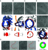
Validation of a Parkin-dependent mitophagy reporter cell line for cryo-FIB milling. Establishment and validation of CLEM mitophagy reporter cells for targeted cryo-FIB milling using mCherry-Parkin and BFP-Mito by confocal microscopy (Scale bar, 10 μm) (A). Parkin recruitment to OA-induced fragmented mitochondria was only observed after dual treatment with OA but not with Oligomycin or Antimycin alone (n = 3 independent fields per replicate per condition) (B). Mitophagy flux assay using the IMM protein Su9 as a probe (C). Quantification of Su9 processing in C (D). Using cryo-fluorescence of BFP-mito to guide the milling process, lamellae targeting the intact (green) or fragmented (magenta) mitochondrial network were generated (E) (Scale bar, 1 μm). (F) Mitochondrial sections observable within OA-treated lamellae were significantly smaller (732 nm average diameter, n = 93) than the healthy untreated network (1,261 nm average diameter, n = 66), (Scale bar, 1 μm).
Fig. 2.
Fig. 2.
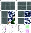
Phagophores target and envelop mitochondrial fragments. An early peanut-shaped double-membrane structure that is likely an early phagophore was identified next to a mitochondrial fragment. Membrane segmentation highlights a slight dimple in this membrane structure that is less than 1 nm deep (A). A larger enveloping double-membrane structure with a 250 nm opening was found targeting a mitochondrial fragment and adjacent to a membrane sheet (B). A fully enveloped mitochondrial fragment in a double-membrane structure that is likely an early mitophagosome with a diameter of approximately 500 nm (black arrows) (C). Stepwise segmentation of the volume in (B) highlighting BLTPs in between the membrane sheet and autophagosomal membranes (black sticks) (D). Two examples of mitochondrial fragments found enveloped in membrane structures consisting of more than two distinct membranes (E) and with membrane segmentation (F). Quantification of phagophore-like structures present in the OA-treated dataset (n = 85 total mitochondrial fragments, n = 71 untargeted and n = 14 phagophore-like) (G). (Scalebar, 100 nm.)
Fig. 3.
Fig. 3.
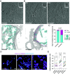
IMS shrinking and decalcification during collapse of the mitochondrial network. Cristae were abundant in untreated mitochondria and still detectable after OA treatment (black asterisks) (A). Segmentation of mitochondrial membranes illustrates cristae abundance and organization in untreated cells, with a sparser distribution following depolarization (B). (C) Quantification of membrane-segmented cristae reveals a significant decrease in average density from 12 to 6 cristae per volume after OA treatment (n = 37 untreated tomograms, n = 81 OA-treated tomograms). (D) Cristae surface area and volume were extracted from membrane segmentations and compared between untreated and OA-treated cells (n = 25 independent cristae per condition, median marked). Unpaired t tests were applied to the averages from all plotted points and used to determine significance. Inspection of tomograms generated from untreated mitochondria revealed abundant matrix-resident electron-dense granules, likely composed of calcium phosphate (black arrows) (E). No such clusters were detected in OA-treated mitochondrial fragments (E). Segmentation of mitochondrial membranes and calcium clusters from tomographic volumes highlights their abundance in the untreated mitochondrial network and absence in depolarized mitochondrial fragments (F). Quantification of calcium clusters reveals an average of 32 clusters were present per untreated tomogram (G). Schematic illustrating mitochondrial IMS shrinking and calcium cluster loss (H). (Scale bar, 100 nm.)
Fig. 4.
Fig. 4.
Mislocalization of ATP synthases to the IBM after OA treatment. In untreated mitochondria, ATP synthases were found in high abundance on cristae membranes and rarely on the IBM (A and Inset). After OA treatment, the OM-associated class of ATP synthases significantly increased in population (B and Insets). Segmentation of the mitochondrial membrane with ATP synthase molecules identified by Pytom back projected onto cristae (cyan spheres) and the OM (magenta spheres) for visualization. Quantification of ATP synthases from each class in untreated (n = 17 IBM, n = 623 cristae) and OA-treated cells (n = 257 IBM, n = 413 cristae) and comparison via Fisher’s exact test with template match density map shown and model docked (RCSB: 8H9T) (C). Live cell Airyscan imaging of mitochondria using TOMM20-mCherry and ATP5F1B-GFP to track ATP synthase localization after depolarization (D). Quantification of localizations in (D) (n = 3 to 4 cells per replicate over three independent replicates with a paired t test used to determine significance) (E). Scale bar, 100 nm unless otherwise indicated.
Fig. 5.
Fig. 5.
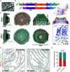
Structural determination of the prohibitin complex and its conformational transition during depolarization. Prohibitin forms a dome-like structure in the IMS of mitochondria (A). Domain architecture for Prohibitin -1 and -2. A heterotetrameric and heterododecameric AlphaFold3 predictions are also shown colored by domains, and the Inset shows the pLDDT scores and secondary structure architecture of the SPFH domain, annotated for Prohibitin -2 (B). (C) Side and top views of EM density maps corresponding to two solved structures of prohibitin complex highlight extra density on the matrix side of the membrane. (D) Back projection of manually picked particles (shown as spheres) onto membrane segmentations from tomograms of both untreated and OA-treated mitochondria (open = salmon, closed = blue). (E) Quantification of prohibitin complexes from each class in untreated (n = 930 open, n = 677 closed) and OA-treated cells (n = 297 open, n = 511 closed) and comparison via Fisher’s exact test. Scale bar, 100 nm unless otherwise indicated.

Update of

References

    1. Uoselis L., Nguyen T. N., Lazarou M., Mitochondrial degradation: Mitophagy and beyond. Mol. Cell 83, 3404–3420 (2023), 10.1016/j.molcel.2023.08.021. - DOI - PubMed
    1. Henrich M. T., Oertel W. H., Surmeier D. J., Geibl F. F., Mitochondrial dysfunction in Parkinson’s disease–A key disease hallmark with therapeutic potential. Mol. Neurodegener. 18, 83 (2023), 10.1186/s13024-023-00676-7. - DOI - PMC - PubMed
    1. Antico O., Thompson P. W., Hertz N. T., Muqit M. M. K., Parton L. E., Targeting mitophagy in neurodegenerative diseases. Nat. Rev. Drug Discov. 24, 276–299 (2025), 10.1038/s41573-024-01105-0. - DOI - PubMed
    1. Clark I. E., et al. , Drosophila pink1 is required for mitochondrial function and interacts genetically with parkin. Nature 441, 1162–1166 (2006), 10.1038/nature04779. - DOI - PubMed
    1. Kitada T., et al. , Mutations in the parkin gene cause autosomal recessive juvenile parkinsonism. Nature 392, 605–608 (1998), 10.1038/33416. - DOI - PubMed

MeSH terms

LinkOut - more resources