. 2025 Sep;12(5):e200455.
doi: 10.1212/NXI.0000000000200455. Epub 2025 Jul 31.

Unknown Immunoregulatory Effects of FcRn Inhibition by Efgartigimod in Myasthenia Gravis: A New Mechanism of Action Beyond IgG Reduction

Affiliations

Unknown Immunoregulatory Effects of FcRn Inhibition by Efgartigimod in Myasthenia Gravis: A New Mechanism of Action Beyond IgG Reduction

Maria Cristina Tarasco et al. Neurol Neuroimmunol Neuroinflamm. 2025 Sep.

Abstract

Background and objectives: Efgartigimod (EFG), a biological drug targeting the IgG recycling neonatal Fc receptor (FcRn), leads to clinical improvements in patients affected by myasthenia gravis (MG), a prototypic autoantibody (Ab)-mediated autoimmune disease affecting neuromuscular junction. Because FcRn is a multifunctional protein expressed in different immune system cells, including B cells, we investigated whether FcRn blockade by EFG may have further immunologic effects, other than IgG reduction, in patients with MG.

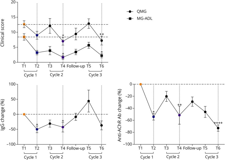
Methods: Anti-acetylcholine receptor Ab-positive (AChR-MG) patients were treated with EFG according to the GENERATIVE protocol. Clinical evaluation, IgG and autoAb quantification, and circulating T-cell and B-cell subpopulation analyses by flow cytometry were performed at different time points. The expression of regulatory plasma cell-related candidate genes (CD38, lymphocyte-activation gene 3 [LAG3], IL-12a, Ebi3) was assessed by real-time PCR in peripheral blood mononuclear cells (PBMCs) from patients on treatment and in PBMCs either untreated or in vitro treated with an EFG-mimicking anti-FcRn monoclonal Ab (mAb) or with EFG (Vyvgart).

Results: A significantly increased percentage of CD19+/CD27+ memory B cells and CD27+/CD138+ plasma cells was observed at the end of EFG treatment cycle 1 and cycle 2 in patients with AChR-MG. Plasma cell increase, maintained up to cycle 3, significantly correlated with Quantitative Myasthenia Gravis score improvement. Moreover, PBMCs from EFG-treated patients showed overexpression of CD38, LAG3, and IL-12a genes, suggesting EFG's ability to induce non-pathogenic regulatory plasma cells. This ability was confirmed in vitro, because anti-FcRn mAb-treated and EFG-treated PBMCs displayed an up-regulation of CD38 and LAG3 compared with untreated cells.

Discussion: Our findings indicate an unknown immunoregulatory action of EFG in patients with AChR-MG, by unraveling a drug effect on B-cell differentiation, and suggest the induction of regulatory plasma cells as a further mechanism, beyond IgG reduction, associated with clinical improvement. A deep understanding of the immunologic effects of EFG can help to optimize its usage over time in individual patients and disclose biomarkers suitable for monitoring the long-term patient-specific response.


Conflict of interest statement

R. Frangiamore has received funding for consulting and speaking from Alexion Pharmaceuticals, UCB, and Argenx. S. Bonanno has received funding for travel, meeting attendance, and advisory board participation from Alexion; Sanofi; Roche; and Biogen. L. Maggi has received funding for travel, meeting attendance, and advisory board participation from Alexion; Sanofi Genzyme; Amicus Therapeutics; Janssen; Argenx; Biogen; Lupin; Roche; patient-centered biopharmaceutical company (PTC) therapeutics; and UCB. F. Vanoli has received funding for travel, meeting attendance, and advisory board participation from Alexion; UCB Pharma; Argenx; and Sanofi. R. Mantegazza has received compensation for participating in advisory boards in relation to MG clinical trial designs, congress participations, and research support from Alexion Pharmaceuticals; ARGENX Pharma; UCB; and PIEM. C. Antozzi has received funding for travel, meeting attendance, and advisory board participation from Alexion; Momenta; Sanofi; Janssen; Argenx; and UCB. P. Cavalcante has received compensation for participating in advisory boards and speaking at scientific meetings from Alexion Pharmaceuticals. The authors are also part of the European Reference Network for rare neuromuscular diseases (ERN-NMD). Go to Neurology.org/NN for full disclosures.

Figures

Figure 1
Figure 1. EFG Treatment Scheme and Time Line of Sample Collection
The protocol of the Expanded Early Access Program (EAP, GENERATIVE) (Version 1.0, September 10, 2021) to EFG consisted of 2 cycles (fixed period) of 4 IV infusions (i.e., one infusion per week), followed by a 30-day follow-up at the end of each cycle. Cycle 3 (i.e., flexible period initiation) started at a variable time interval from cycle 2 end (3.1 ± 2.7 months), depending on disease worsening. Blood samples for serum and peripheral blood mononuclear cell isolation were collected from patients with MG at the following time points: baseline or T1, i.e., before the 1st infusion of cycle 1; T2, i.e., before the 4th infusion of cycle 1; T3, i.e., before the 1st infusion of cycle 2; T4, i.e., before the 4th infusion of cycle 2; follow-up, i.e., at the follow-up visit after cycle 2 and performed at 30 days after the end of the cycle; T5, i.e., before the 1st infusion of cycle 3; and T6, i.e., before the 4th infusion of cycle 3. EFG = efgartigimod; GENERATIVE = GENERalized myAsThenia graVis Efgartgimod; MG = myasthenia gravis.
Figure 2
Figure 2. Clinical Improvement in Patients With AChR-MG Treated With EFG
QMG and MG-ADL scores and percentage of total IgG and anti-AChR antibody changes in EFG-treated patients with AChR-MG (n = 9). In the graphs, the mean values ± SEM calculated for each measure in all the patients at each time point are shown. *p < 0.05, **p < 0.01, ****p < 0.0001, Friedman test with Dunn multiple comparison tests. AChR-MG = anti-acetylcholine receptor Ab–positive; EFG = efgartigimod; MG-ADL = Myasthenia Gravis Activities of Daily Living; QMG = Quantitative Myasthenia Gravis.
Figure 3
Figure 3. Increase in Memory B Cells and Plasma Cells in EFG-Treated Patients With AChR-MG
Results of B-cell subpopulation analysis by flow cytometry in longitudinally collected PBMCs from patients with AChR-MG during EFG therapy. For T5 and T6, the analyses included 6 of the 9 patients. At each time point, the mean values ±SEM of the percentage of the following B-cell subpopulations have been reported: (1) total B cells as CD19+ cells, (2) naïve B cells as CD19+/CD27- cells, (3) memory B cells as CD19+/CD27+ cells, and (4) plasma cells as CD27+/CD138+ cells. A significant increase in the percentage of memory B cells was observed at T2 and of plasma cells at both T4 and T6 compared with the baseline. *p < 0.05, Kruskal-Wallis with uncorrected Dunn test. AChR-MG = anti-acetylcholine receptor Ab–positive; EFG = efgartigimod; PBMC = peripheral blood mononuclear cell.
Figure 4
Figure 4. Changes in Memory B Cells and Plasma Cells in Individual Patients and Correlation With Clinical Improvement and Increased Expression of Regulatory Plasma Cell Markers in EFG-Treated Patients With AChR-MG
(A) Graphs showing the intra-patient behavior of CD19+/CD27+ memory B cells and CD138+/CD27+ plasma cells at T2 and T4, respectively, compared with the baseline (T1). *p < 0.05, paired t test. (B) Significant negative correlation estimated by Spearman correlation analysis (r: −0.81, p < 0.05) between QMG score changes (T4 vs T1) and the plasma cell increase (T4/T1) in patients with AChR-MG showing an increase in these cells at T4. (C and D) Real-time PCR analysis showing transcriptional levels of CD38 (C) and LAG3, IL-12a, and Ebi3 (D) genes in PBMCs of patients with AChR-MG (n = 8) before (T1) and on treatment with EFG (T4, i.e., 4th infusion of cycle 2). An upregulation of all genes was found at T4 compared with T1. In the “before-after” graph, data are expressed as relative expression values (2−∆Ct × 100) normalized against the endogenous control 18S. The line in red indicates a non-responder patient who did not undergo improvement on EFG therapy. The line in blue indicates a patient, initially a responder, but then requiring to begin cycle 3 of the drug earlier than the other patients. Differences between the 2 time points were significant for LAG3 after excluding the non-responder patient from the analyses (not shown). *p < 0.05, Wilcoxon matched-pairs signed-rank test. A significant positive correlation (r: 0.84; p < 0.01) was found between CD138+/CD27+ plasma cell percentage and transcriptional levels of CD38 at T4 by the Pearson correlation test (C). AChR-MG = anti-acetylcholine receptor Ab–positive; EFG = efgartigimod; PBMC = peripheral blood mononuclear cell.
Figure 5
Figure 5. FcRn Expression in B Cells and In Vitro Effect of Anti-FcRn Treatment
(A) Double immunofluorescence staining showing the expression of FcRn (green) in CD20+ (red) B cells of MG PBMCs collected at T1 (baseline). Magnification bars: 10 µm. (B) Representative overlay histograms of flow cytometry analysis performed to assess percentage of CD19+/CD27+ B cells in PBMCs from healthy controls (n = 2) and treatment-naïve patients with MG (n = 2) after 24-hour treatments performed in triplicate with (1) EFG (Vyvgart; dose I: 1 µg/mL) or human IgG1 as negative control and (2) an anti-FcRn mAb (sc-271745, Santa Cruz Biotechnology; 1 µg/mL) or mouse IgG2a as isotype control. Basal: untreated cells; Neg Control: unstained cells. EFG = efgartigimod; PBMC = peripheral blood mononuclear cell.
Figure 6
Figure 6. In Vitro Effects of Anti-FcRn Treatment on B-Cell Frequency and Expression of CD38 and LAG3
(A) Percentage of CD19+ B cells measured by flow cytometry in PBMCs from healthy controls (CTR, n = 2, white dots) and treatment-naïve patients with MG (n = 2, black dots), after 24-hour treatments performed in triplicate with EFG (Vyvgart; dose I: 1 µg/mL) or human IgG1 as negative control and (2) an anti-FcRn mAb (sc-271745, Santa Cruz Biotechnology; 1 µg/mL) or mouse IgG2a as an isotype control. Data are expressed as mean ± SEM of percentage changes in treated PBMCs relative to untreated cells. An outlier value (−85%) in the group of IgG1-treated cells was excluded from the analyses, according to the Grubbs test. (B) Real-time PCR analysis of transcriptional levels of CD38 and LAG3 in PBMCs from healthy controls (CTR, n = 1, white dots) and treatment-naïve patients with MG (n = 2, black dots), after 24-hour treatments performed in triplicate with EFG or the anti-FcRn mAb and their relative controls. Data are presented as mean ± SEM of 2−∆Ct × 100 relative expression values normalized with the endogenous control 18S. *p < 0.05, ***p < 0.01, **p < 0.01, ***p < 0.001, the Friedman test with Dunn's multiple comparison tests. MG = myasthenia gravis; PBMC = peripheral blood mononuclear cell.

References

    1. Kaminski HJ, Sikorski P, Coronel SI, Kusner LL. Myasthenia gravis: the future is here. J Clin Invest. 2024;134(12):e179742. doi: 10.1172/JCI179742 - DOI - PMC - PubMed
    1. Lazaridis K, Tzartos SJ. Myasthenia gravis: autoantibody specificities in myasthenia gravis; implications for improved diagnostics and therapeutics. Front Immunol. 2020;11:212. doi: 10.3389/fimmu.2020.00212 - DOI - PMC - PubMed
    1. Narayanaswami P, Sanders DB, Wolfe G, et al. International consensus guidance for management of myasthenia gravis: 2020 update. Neurology. 2021;96(3):114-122. doi: 10.1212/WNL.0000000000011124 - DOI - PMC - PubMed
    1. Mantegazza R, Antozzi C. When myasthenia gravis is deemed refractory: clinical signposts and treatment strategies. Ther Adv Neurol Disord. 2018;11:1756285617749134. doi: 10.1177/1756285617749134 - DOI - PMC - PubMed
    1. Cortés-Vicente E, Álvarez-Velasco R, Paradas C, et al. Drug‐refractory myasthenia gravis: clinical characteristics, treatments, and outcome. Ann Clin Transl Neurol. 2022;9(2):122-131. doi: 10.1002/acn3.51492 - DOI - PMC - PubMed