

Integrative Analysis of Novel Ferroptosis-Related Genes Signatures as Prognostic Biomarkers in Ovarian Cancer
- PMID: 40744688
- PMCID: PMC12313356
- DOI: 10.1002/cnr2.70284
Integrative Analysis of Novel Ferroptosis-Related Genes Signatures as Prognostic Biomarkers in Ovarian Cancer
Abstract
Background: Ferroptosis, an iron-dependent form of cell death, has been implicated in the pathogenesis of several types of cancer. Nevertheless, the exact correlation between ferroptosis-related gene mutations and their influence on ovarian cancer (OV) diagnosis and treatment strategies remains to be fully elucidated. It is crucial to identify the ferroptosis-related gene signature in OV and elucidate the impact of these mutations and their expression on the diagnosis and treatment of OV.
Methods: In this study, we collected data from the TCGA and GEO databases. We utilized various tools and packages for data analysis, including the cBio Cancer Genomics Portal, Tumor Immune Estimation Resource (TIMER), GSVA package, and WGCNA R packages.
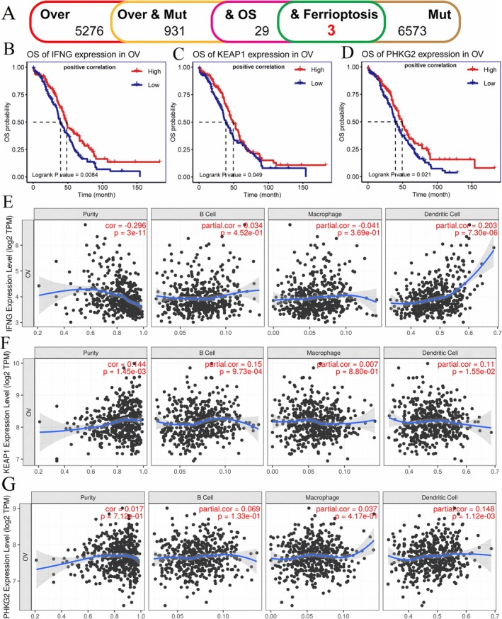
Results: Our results showed that ferroptosis subtypes 1 (FS1) and 2 (FS2) exhibited different levels of expression and tumor mutation burden (TMB). FS2 had a higher TMB level and survival rate compared to FS1. Furthermore, our analysis identified three ferroptosis-related genes, including IFNG, KEAP1, and PHKG2, as key biomarkers in prognosis prediction and potential targets for OV cancer therapy. The elevated expression levels of IFNG, KEAP1, and PHKG2 were found to be correlated with a good prognosis. These three genes showed a positive correlation with TMB in OV. We also observed that high TMB was more robustly associated with immune response-related gene expression, including CD28, CD40L, and type I IFN family members. Moreover, high TMB was associated with increased T cell infiltration and exhibited a distinct gene signature, which highlights the potential of IFNG, KEAP1, and PHKG2 as predictive markers for T cell infiltration and the tumor microenvironment status in OV. A significant correlation exists between the expression levels of KEAP1 and PHKG2 and TMB in OV cell lines.
Conclusion: In conclusion, our study identified KEAP1, IFNG, and PHKG2 as potential prognostic biomarkers and therapeutic targets in OV. Their expression and mutation burden were correlated with a good prognosis. The association between ferroptosis subtypes, TMB, and survival rates further supports the relevance of these biomarkers. Additionally, the positive correlation between KEAP1, IFNG, and PHKG2 with TMB and immune response-related gene expression highlights their potential as predictive markers for immunotherapy efficacy in OV. The observed association of high TMB with increased T cell infiltration and distinct gene signature further emphasizes its role as a potential biomarker for immune response. Further research is warranted to validate these findings and explore their clinical implications in OV treatment.
Keywords: ferroptosis; immunotherapy; ovarian cancer; tumor microenvironment; tumor mutation burden.
© 2025 The Author(s). Cancer Reports published by Wiley Periodicals LLC.
Conflict of interest statement
The authors declare no conflicts of interest.
Figures


Similar articles
-
Interplay between tumor mutation burden and the tumor microenvironment predicts the prognosis of pan-cancer anti-PD-1/PD-L1 therapy.Front Immunol. 2025 Jul 24;16:1557461. doi: 10.3389/fimmu.2025.1557461. eCollection 2025. Front Immunol. 2025. PMID: 40777041 Free PMC article.
-
Ferroptosis-related LINC02535/has-miR-30c-5p/EIF2S1 axis as a novel prognostic biomarker involved in immune infiltration and progression of PDAC.Cell Signal. 2024 Nov;123:111338. doi: 10.1016/j.cellsig.2024.111338. Epub 2024 Aug 6. Cell Signal. 2024. PMID: 39117252
-
Comprehensive pan-cancer analysis reveals NTN1 as an immune infiltrate risk factor and its potential prognostic value in SKCM.Sci Rep. 2025 Jan 25;15(1):3223. doi: 10.1038/s41598-025-85444-x. Sci Rep. 2025. PMID: 39863609 Free PMC article.
-
Impact of residual disease as a prognostic factor for survival in women with advanced epithelial ovarian cancer after primary surgery.Cochrane Database Syst Rev. 2022 Sep 26;9(9):CD015048. doi: 10.1002/14651858.CD015048.pub2. Cochrane Database Syst Rev. 2022. PMID: 36161421 Free PMC article.
-
Systemic pharmacological treatments for chronic plaque psoriasis: a network meta-analysis.Cochrane Database Syst Rev. 2021 Apr 19;4(4):CD011535. doi: 10.1002/14651858.CD011535.pub4. Cochrane Database Syst Rev. 2021. Update in: Cochrane Database Syst Rev. 2022 May 23;5:CD011535. doi: 10.1002/14651858.CD011535.pub5. PMID: 33871055 Free PMC article. Updated.
References
-
- Konstantinopoulos P. A. and Matulonis U. A., “Clinical and Translational Advances in Ovarian Cancer Therapy,” Nature Cancer 4 (2023): 1239–1257. - PubMed
-
- Li B., Yang L., Peng X., et al., “Emerging Mechanisms and Applications of Ferroptosis in the Treatment of Resistant Cancers,” Biomedicine & Pharmacotherapy 130 (2020): 110710. - PubMed
MeSH terms
Substances
Grants and funding
LinkOut - more resources
Full Text Sources
Medical
