Review
. 2025 Aug 1;11(1):358.
doi: 10.1038/s41420-025-02654-y.

Redox mechanism of glycerophospholipids and relevant targeted therapy in ferroptosis

Affiliations
Review

Redox mechanism of glycerophospholipids and relevant targeted therapy in ferroptosis

Shuwei Chang et al. Cell Death Discov. .

Abstract

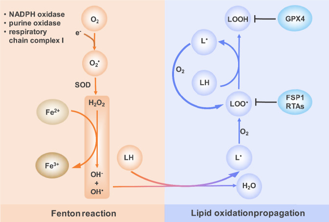
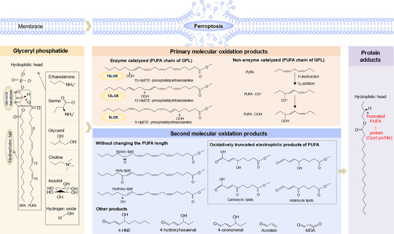
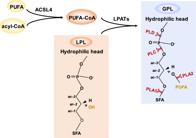
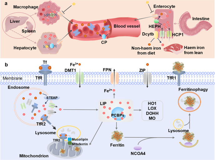
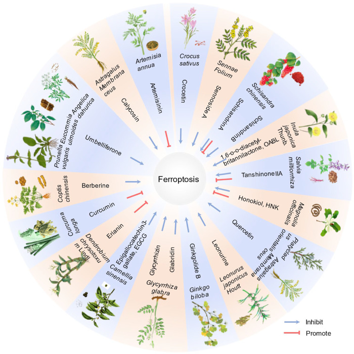
Ferroptosis, an iron-dependent form of regulated cell death driven by redox dysregulation, is defined by iron overload, reactive oxygen species overproduction, and subsequent peroxidation of polyunsaturated fatty acid-containing phospholipids, notably glycerophospholipids. This review comprehensively delineates the enzymatic such as lipoxygenases and non-enzymatic including Fenton reaction pathways governing glycerophospholipid peroxidation. Furthermore, we systematically dissect fine regulation of iron ions, including absorption, transport, and redox state transition. Given pathophysiological relevance of ferroptosis to numerous diseases, especially neurodegenerative disorders and various cancers, we evaluate emerging therapeutic strategies targeting key ferroptosis nodes, with a primary focus on the key enzymes involved in lipid peroxidation, transferrin receptor-mediated endocytosis mechanism and traditional Chinese medicine. Our work provides a direction for advancing ferroptosis research and developing combinatorial therapies that synergize ferroptosis induction with conventional treatments.


Conflict of interest statement

Competing interests: The authors declare no competing interests.

Figures

Fig. 1
Fig. 1. Mechanism of lipid peroxidation.
Superoxide anion radicals are enzymatically produced by sources such as NADPH oxidase, purine oxidase, mitochondrial complex I etc. These radicals are then converted to hydrogen peroxide (H₂O₂) by superoxide dismutase (SOD). H₂O₂ reacts with Fe²⁺, oxidizing it to Fe³⁺ and producing the highly reactive hydroxyl radical (HO•) via the Fenton reaction. Hydroxyl radicals abstract hydrogen atoms from oxidizable molecules like polyunsaturated lipids (LH), yielding carbon-centered lipid radicals (L•). These rapidly react with oxygen (O₂) to form lipid peroxyl radicals (LOO•). LOO• propagate lipid oxidation by abstracting a hydrogen atom from adjacent polyunsaturated lipids (LH). This forms lipid hydroperoxides (LOOH) and generates a new lipid radical (L•), which rapidly reacts with oxygen to yield another peroxyl radical (LOO•), sustaining the chain reaction. Inhibiter of ferroptosis: GPX4, FSP1, RTAs. GPX4, glutathione peroxidase 4; FSP1, ferroptosis suppressor protein-1; RTAs, radical-trapping antioxidants.
Fig. 2
Fig. 2. Peroxidation mechanisms of glycerophospholipids containing PUFA.
Glycerol serves as the backbone of phospholipids, with two FA chains esterified at the sn-1 and sn-2 positions, and a polar head group attached at the sn-3 position. Common polar head groups in phospholipids include ethanolamine, serine, glycerol, choline, and inositol. PUFAs are typically esterified at the sn-2 position of phospholipids. Their structure, featuring two or more cis-configured double bonds, creates bis-allylic hydrogen atoms with low bond dissociation energies, making these sites highly susceptible to hydrogen abstraction. LP PUFAs undergo oxidation via both enzymatic (e.g., LOXs) and non-enzymatic ways, forming primary oxidized products. These primary products can be further redox to form the second products with intact FA chains (e.g., epoxy-, keto-, hydroxy-containing PLs). Or the electrophilic, chain-shortened products (e.g., carboxy, aldehyde relative PLs) due to the low dissociation energy of the O–O bond. Key byproducts of this process, notably 4-HNE and MDA (middle), readily form covalent adducts with nucleophilic residues on proteins (e.g., cysteine, histidine, and lysine), altering protein function and contributing to oxidative stress pathology (right). PUFA, polyunsaturated fatty acyl; GPL, glycerophospholipid; HpETE, 6 hloride 6 d 6 l-eicosatetraenoic acid; 4-HNE, 4-hydroxynonenal; MDA, malondialdehyde.
Fig. 3
Fig. 3. Glycerophospholipid (GPL) regulation.
ACSL4 catalyzes PUFA and acyl-CoA to form PUFA-CoA, which then forms GPL with LPL in the presence of LPAAT enzyme. GPLs is hydrolyzed by phospholipases at specific sites: PLA1 cleaves the sn-1 fatty acyl ester, PLA2 the sn-2 fatty acyl ester, PLC the phosphodiester bond between glycerol and the phosphate group, and PLD the phosphodiester bond between phosphate and the head group. PUFA polyunsaturated fatty acid, ACSL4 acyl-CoA synthesis long-chain family member, LPL lysophospholipid, LPLATs lysophospholipid acyltransferases, GPL glycerophospholipid, SFA saturated fatty acid.
Fig. 4
Fig. 4. Irons homeostasis regulation.
a Systemic iron flux. Macrophages in the liver and spleen take up and digest senescent red blood cells, releasing the iron into the plasma; the remainder comes from dietary sources. Dietary iron comprises heme iron (from lean) and non-heme iron. Intestinal enterocytes absorb heme iron via HCP1, while non-heme iron is absorbed by DMT1 after being reduced to Fe²⁺ by Dcytb, which is primarily localized in the enterocyte’s apical membrane. FPN, the only known iron ions exporter, which mediates Fe2+ export in mammalian cells. Multicopper oxidases oxidize Fe²⁺ to Fe³⁺. HEPH (highly expressed in intestines) and CP (hepatocyte-derived, circulating in plasma) both exhibit ferroxidase activity. HEPH presumably works in conjunction with FPN. b Cellular iron regulation. Tf binds Fe³⁺ and delivers it to cells via TfR-mediated endocytosis, forming endosomes. Within endosomes, Fe³⁺ dissociates from Tf, is reduced to Fe²⁺ by STEAP3, and is transported into the cytosol by DMT1 or ZIP transporters. Human TfR1 was found belonging to cell surface receptor for H ferritin. TfR2 contains mitochondrial targeting motif, facilitates irons delivery to the mitochondria; Lysosomes containing TfR2-α translocate to mitochondria and dock with them, facilitating iron transfer. Cytosolic labile iron is stored within ferritin cages with the assistance of PCBPs. During iron deficiency, ferritin interacts with the receptor NCOA4 and is degraded via ferritinophagy. Furthermore, PCBPs assist in other cellular iron processes: transferring iron from the importer DMT1, exporting iron via FPN, degrading heme through HO1; and regulating iron-dependent enzymes involved in lipid peroxidation (LOXs); other enzymes including DOHH, MO or enzymes in mitochondria. Tf transferrin, TfR transferrin receptor, FPN ferroportin, DMT1 divalent metal transporter 1, Dcytb duodenal cytochrome b, ZIP ZRT/IRT-like protein, STEAP six-transmembrane epithelial antigen of the prostate. HCP haem carrier protein 1, CP Ceruloplasmin, HEPH Hephaestin, LIP Labile ion pool, PCBPs (poly(Rc)-binding proteins 1-4), HO1 heme degradation via heme oxygenase 1, LOXs lipoxygenases, DOHH deoxyhypusine hydroxylase, MO monooxygenases.
Fig. 5
Fig. 5. Pharmaceutical ingredients function of TCM in ferroptosis.
This figure summarizes 19 traditional Chinese herbs and their associated bioactive compounds, with documented effects on ferroptosis regulation based on current research. Note: As research advances, additional bioactive compounds and regulatory functions in ferroptosis are anticipated to be identified. This diagram should be updated accordingly to reflect new evidence.

Similar articles

References

    1. Dixon SJ, Lemberg KM, Lamprecht MR, Skouta R, Zaitsev EM, Gleason CE, et al. Ferroptosis: an iron-dependent form of nonapoptotic cell death. Cell. 2012;149:1060–72. - PMC - PubMed
    1. Li X, He J, Gao X, Zheng G, Chen C, Chen Y, et al. GPX4 restricts ferroptosis of NKp46+ILC3s to control intestinal inflammation. Cell Death Dis. 2024;15:687. - PMC - PubMed
    1. Chen Y, Fang Z-M, Yi X, Wei X, Jiang D-S. The interaction between ferroptosis and inflammatory signaling pathways. Cell Death Dis. 2023;14:205. - PMC - PubMed
    1. Yuan Y, Mei Z, Qu Z, Li G, Yu S, Liu Y, et al. Exosomes secreted from cardiomyocytes suppress the sensitivity of tumor ferroptosis in ischemic heart failure. Signal Transduct. Targeted Ther. 2023;8:121. - PMC - PubMed
    1. Fang X, Ardehali H, Min J, Wang F. The molecular and metabolic landscape of iron and ferroptosis in cardiovascular disease. Nat Rev Cardiol. 2022;20:7–23. - PMC - PubMed

LinkOut - more resources