. 2025 Jul 31;15(1):27975.
doi: 10.1038/s41598-025-08642-7.

Enhancement of angiotensin II type 1 receptor-associated protein suppresses kidney inflammation in a mouse model of aristolochic acid nephropathy

Affiliations

Enhancement of angiotensin II type 1 receptor-associated protein suppresses kidney inflammation in a mouse model of aristolochic acid nephropathy

Rika Furuta et al. Sci Rep. .

Abstract

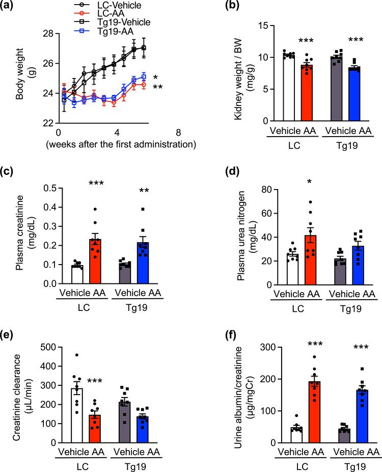
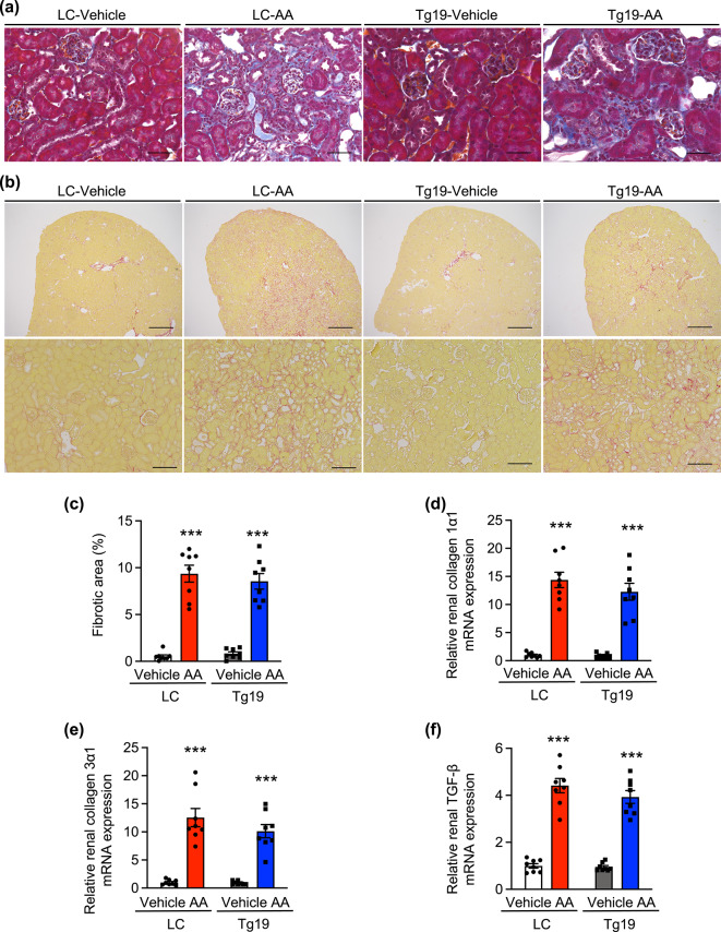
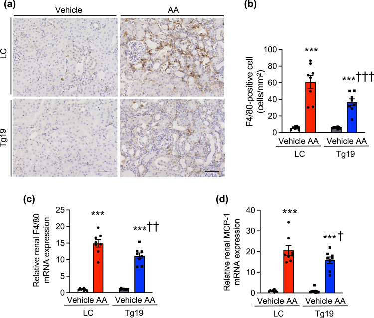
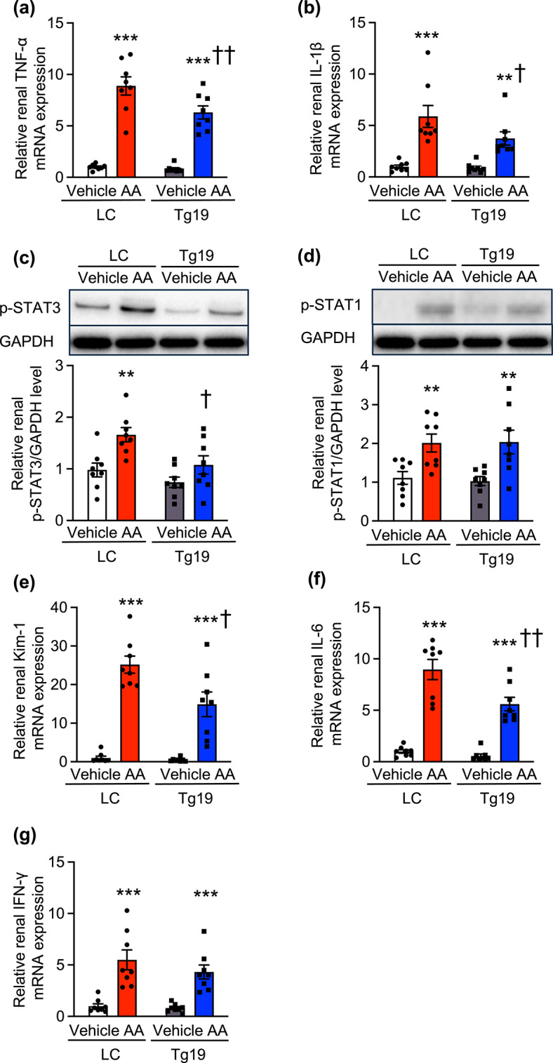
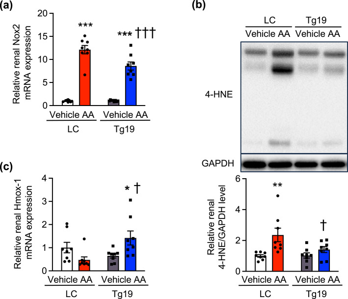
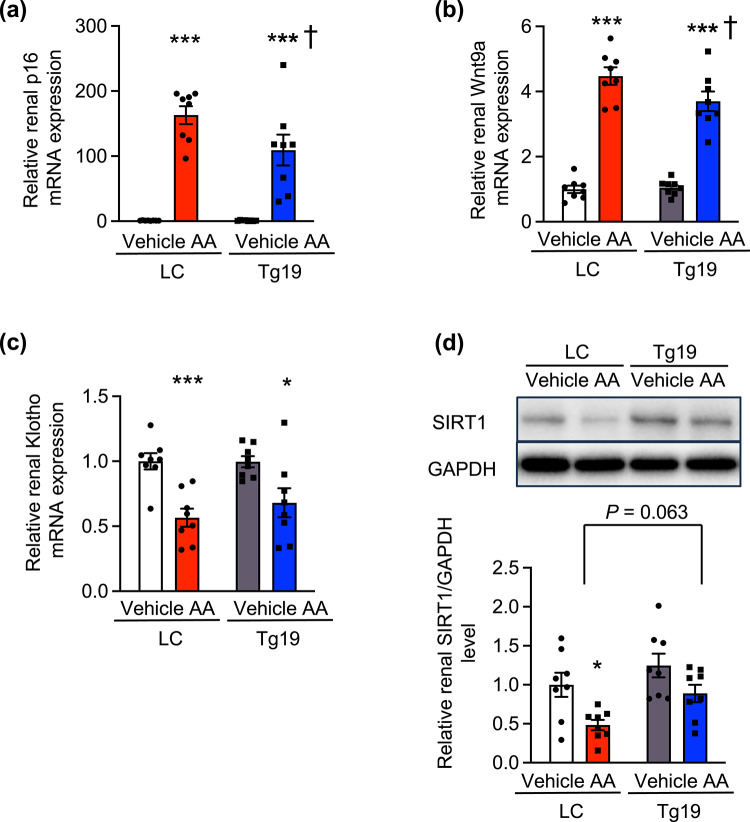
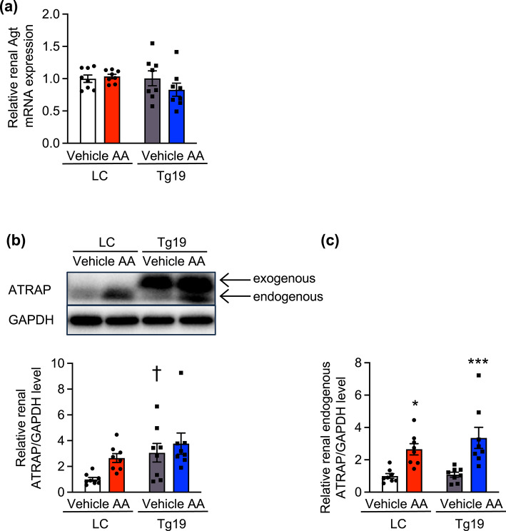
Chronic kidney disease generally progresses to irreversible fibrosis through chronic inflammation and age-related changes. We had previously reported that genetic knockdown of angiotensin II type 1 receptor (AT1R)-associated protein (ATRAP) exacerbates aging-associated kidney tubulointerstitial fibrosis in mice. However, whether enhanced ATRAP expression can suppress renal fibrosis and senescence in vivo remains unknown. Recently, we proposed that aristolochic acid nephropathy (AAN) could be used for modeling kidney aging with fibrosis. The present study aimed to investigate the functional role of ATRAP in aging-associated kidney fibrosis and inflammation using ATRAP transgenic (Tg19) mice subjected to AAN. AA administration caused histological renal fibrosis and enhanced ATRAP expression had no apparent effect on AA-induced renal fibrosis. However, enhanced ATRAP expression significantly suppressed AA-induced macrophage infiltration concomitant with reductions in inflammation-related, macrophage-related and senescence-related gene expression in the kidneys. Furthermore, the renal expression of anti-aging gene (Klotho, Sirtuin1) was significantly reduced in control mice in response to AA administration, whereas AA-mediated downregulation of Sirtuin1 expression was tendentially less prominent in Tg19 mice. Collectively, the enhancement of ATRAP expression failed to ameliorate renal fibrosis but partially attenuated renal inflammation and cellular senescence in AAN. Thus, ATRAP is a potential therapeutic target against renal inflammation and senescence.

Keywords: Angiotensin II type 1 receptor-associated protein; Anti-aging genes; Aristolochic acid nephropathy; Chronic kidney disease; Renal fibrosis.


Conflict of interest statement

Declarations. Competing interests: The authors declare no competing interests.

Figures

Fig. 1
Fig. 1
Effect of AA treatment on body weight change, tissue weight, renal function, and urinary albumin excretion in ATRAP transgenic (Tg19) and littermate control (LC) mice. (a) BW changes, (b) kidney weight/BW ratios, (c) plasma creatinine level, (d) plasma urea nitrogen level, (e) creatinine clearance, and (f) urinary albumin/creatinine ratio in the LC and Tg19 mice at 8 weeks after vehicle or AA administration. Values are expressed as mean ± SE (n = 8 per group) and analyzed using two-way ANOVA with Bonferroni’s post-hoc test. *P < 0.05 vs. vehicle; **P < 0.01 vs. vehicle; ***P < 0.001 vs. vehicle. AA, aristolochic acid; BW, body weight.
Fig. 2
Fig. 2
Effects of AA treatment on renal histological fibrosis and fibrosis-related gene expression in ATRAP transgenic (Tg19) and littermate control (LC) mi. (a) Representative micrographs of Masson’s trichrome-stained renal sections from LC and Tg19 mice. Magnification ×400 (bar: 50 µm). (b) Representative micrographs of picrosirius red-stained renal sections from LC and Tg19 mice. Upper panel, magnification ×40 (bar: 500 µm), lower panel, magnification ×200 (bar: 100 µm). (c) Quantitative analysis of kidney fibrotic area (n = 8 per group). Relative renal mRNA expression of (d) collagen 1α1, (e) collagen 3α1, and (f) TGF-β in the LC and Tg19 mice. Values are expressed as mean ± SE (n = 8 per group) and analyzed using two-way ANOVA with Bonferroni’s post-hoc test. ***P < 0.001 vs. vehicle. AA, aristolochic acid; TGF-β, transforming growth factor-β.
Fig. 3
Fig. 3
Effects of AA treatment on renal histological inflammation and macrophage-related gene expression in ATRAP transgenic (Tg19) and littermate control (LC) mice. (a) Representative micrographs of anti-F4/80-stained renal sections from LC and Tg19 mice. Magnification ×400 (bar: 50 µm). (b) Number of F4/80-positive cells in the kidneys of LC and Tg19 mice. Relative renal mRNA expression of (c) F4/80 and (d) MCP-1 in the LC and Tg19 mice. Values are expressed as mean ± SE (n = 8 per group) and were analyzed using two-way ANOVA with Bonferroni’s post-hoc test. ***P < 0.001 vs. vehicle; P < 0.05 vs. LC mice; ††P < 0.01 vs. LC mice; †††P < 0.001 vs. LC mice. AA, aristolochic acid; MCP-1, monocyte chemoattractant protein 1.
Fig. 4
Fig. 4
Effects of AA treatment on inflammation-related gene expression in ATRAP transgenic (Tg19) and littermate control (LC) mice. Relative renal mRNA expression of (a) TNF-α, (b) IL-1, (e) Kim-1, (f) IL-6 and (g) IFN-γ, and protein expression of (c) p-STAT3 and (d) p-STAT1 in the LC and Tg19 mice. Values are expressed as mean ± SE (n = 8 per group) and were analyzed using two-way ANOVA with Bonferroni’s post-hoc test. **P < 0.01 vs. vehicle; ***P < 0.001 vs. vehicle; P < 0.05 vs. LC mice; ††P < 0.01 vs. LC mice. AA, aristolochic acid; TNF-α, tumor necrosis factor-alpha; IL-1β, interleukin 1β; Kim-1, Kidney injury molecule 1; IL-6, interleukin 6; IFN-γ, interferon-γ; p-STAT3, phosphorylated signal transducer and activator of transcription 3; GAPDH, glyceraldehyde-3-phosphate dehydrogenase; p-STAT1, phosphorylated signal transducer and activator of transcription 1.
Fig. 5
Fig. 5
Effects of AA treatment on renal expression of cellular oxidative stress-related genes in ATRAP transgenic (Tg19) and litter mate control (LC) mice. Relative renal mRNA expression of (a) Nox2, (c) Hmox-1, and (b) protein expression of 4-HNE in the LC and Tg19 mice. Values are expressed as mean ± SE (n = 8 per group) and were analyzed using two-way ANOVA with Bonferroni’s post-hoc test. **P < 0.01 vs. vehicle; ***P < 0.001 vs. vehicle; P < 0.05 vs. LC mice; †††P < 0.001 vs. LC mice. AA, aristolochic acid; Nox2, a component of nicotinamide adenine dinucleotide phosphate oxidase2; 4-HNE, 4-Hydroxynonenal; GAPDH, glyceraldehyde-3-phosphate dehydrogenase; Hmox-1, Heme oxygenase 1.
Fig. 6
Fig. 6
Effects of AA treatment on renal expression of cellular senescence-related genes in ATRAP transgenic (Tg19) and littermate control (LC) mice. Relative renal mRNA expression of (a) p16, (b) Wnt9a and (c) Klotho, and (d) protein expression of SIRT1 in LC and Tg19 mice. Values are expressed as mean ± SE (n = 8 per group) and were analyzed using two-way ANOVA with Bonferroni’s post-hoc test. *P < 0.05 vs. vehicle; ***P < 0.001 vs. vehicle; P < 0.05 vs. LC mice. Wnt9a, Wnt family member 9A; SIRT1, Sirtuin1; AA, aristolochic acid; GAPDH, glyceraldehyde-3-phosphate dehydrogenase.
Fig. 7
Fig. 7
Effect of AA treatment on Agt and ATRAP expression in the kidneys of ATRAP transgenic (Tg19) and littermate control (LC) mice. (a) Relative renal mRNA expression of Agt in the LC and Tg19 mice. Relative renal protein expression of (b) ATRAP and (c) endogenous ATRAP. Values are expressed as mean ± SE (n = 8 per group) and analyzed using two-way ANOVA with Bonferroni’s post-hoc test. *P < 0.05 vs. vehicle; ***P < 0.001 vs. vehicle; P < 0.05 vs. LC mice. AA, aristolochic acid; GAPDH, glyceraldehyde-3-phosphate dehydrogenase; Agt, angiotensinogen; ATRAP, angiotensin II type 1 receptor-associated protein.

Similar articles

References

    1. O’Sullivan, E. D., Hughes, J. & Ferenbach, D. A. Renal aging: Causes and consequences. J. Am. Soc. Nephrol.28(2), 407–420 (2017). - PMC - PubMed
    1. Childs, B. G., Durik, M., Baker, D. J. & van Deursen, J. M. Cellular senescence in aging and age-related disease: From mechanisms to therapy. Nat. Med.21(12), 1424–1435 (2015). - PMC - PubMed
    1. Zhou, B. et al. The emerging role of cellular senescence in renal diseases. J. Cell Mol. Med.24(3), 2087–2097 (2020). - PMC - PubMed
    1. Cao, Q., Harris, D. C. & Wang, Y. Macrophages in kidney injury, inflammation, and fibrosis. Physiology30(3), 183–194 (2015). - PubMed
    1. Feng, Y. et al. Wnt/ β-catenin-promoted macrophage alternative activation contributes to kidney fibrosis. J. Am. Soc. Nephrol.29(1), 182–193 (2018). - PMC - PubMed

MeSH terms

LinkOut - more resources