. 2025 Jul 31;15(1):28039.
doi: 10.1038/s41598-025-12444-2.

Fusion of SARS-CoV-2 neutralizing LCB1 peptide with Bacillus amyloliquefaciens RNase improves antiviral efficacy

Affiliations

Fusion of SARS-CoV-2 neutralizing LCB1 peptide with Bacillus amyloliquefaciens RNase improves antiviral efficacy

Nikita N Kostin et al. Sci Rep. .

Abstract

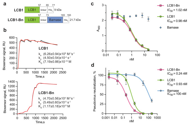
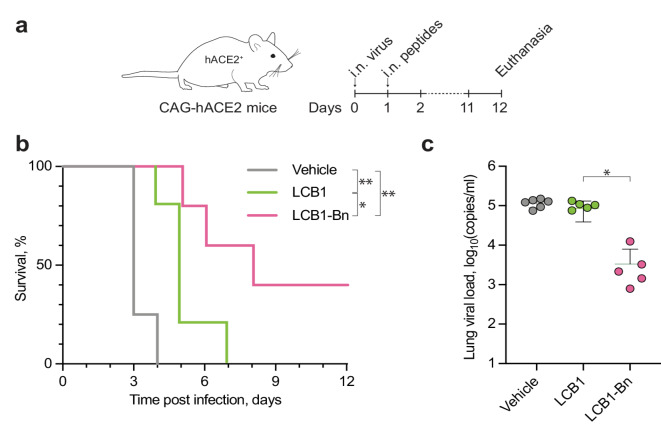
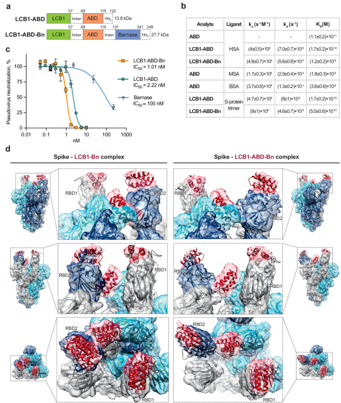
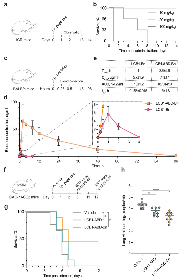
Virus-neutralizing peptides (VNPs) emerged as promising antiviral drug candidates with unprecedented specificity and cost-effectiveness during the recent COVID-19 pandemic. However, limited avidity, lack of effector functions, short circulatory half-life, and restricted administration routes make them inferior compared to neutralizing antibodies. To address these constraints, a potent VNP that targets the SARS-CoV-2 S protein is combined with Barnase, a highly active RNA-cleaving enzyme from Bacillus amyloliquefaciens. The resulting LCB1-Barnase (LCB1-Bn) chimera retains strong binding affinity for the SARS-CoV-2 S protein and demonstrates a fourfold reduction in IC50 compared to the LCB1 peptide alone in competitive ELISA and in in vitro neutralization tests. In transgenic CAG-hACE2 mice infected with wild-type SARS-CoV-2, intranasal administration of LCB1-Bn significantly improves survival and reduces viral load by 29-fold. To extend circulation life and allow systemic intravenous administration, an albumin-binding domain (ABD) from Streptococcus protein G is added to LCB1-Bn, producing LCB1-ABD-Bn fusion protein which displays a 95-fold increase in serum half-life. LCB1-ABD-Bn exhibits good tolerability at doses below 10 mg/kg and provides protection of SARS-CoV-2-infected CAG-hACE2 animals in 24-hour post-infection intraperitoneal treatment. Cryo-EM reveals the LCB1-ABD-Bn's tight interaction with S protein RBD domains, highlighting its potential as a promising drug candidate against SARS-CoV-2.

Keywords: Antiviral peptide; Barnase; Half-life extension; Pharmacokinetics; SARS-CoV-2; Virus neutralizing peptides (VNPs).


Conflict of interest statement

Declarations. Competing interests: The authors declare no competing interests.

Figures

Fig. 1
Fig. 1
Functional characterization of LCB1 and its conjugate with Barnase in vitro. (a) Design of the polypeptides: two variants were expressed, a His-tagged LCB1 and a chimeric version with Barnase fused at the C-terminus, referred to as LCB1-Bn. (b) Single-cycle kinetic SPR analysis of LCB1 and LCB1-Bn binding to the WT trimeric S protein. Polypeptides were applied successively at 25, 50, 100, 200, and 400 nM. Red curves represent the raw data while black curves are a 1:1 binding model fitting. ka – association rate, kd—dissociation rate, KD - affinity constant; RU, response units. (c) Competitive ELISA of the polypeptides binding with the S protein in the presence of ACE2. Mean values ± SD are plotted as curves. (d) Virus-neutralizing activity of polypeptides in the pseudovirus system. Mean values ± SD are plotted as curves.
Fig. 2
Fig. 2
Barnase enhances the protective properties of LCB1 peptide in vivo in transgenic CAG-hACE2 mice infected with WT SARS-CoV-2 virus. (a) Transgenic CAG-hACE2 mice were exposed to intranasal infection (i.n.) with the live virus. The next day, the polypeptides were administered i.n. Survival was monitored over a period of 12 d, the mice were euthanized, and the viral load was determined in the lungs. (b) Survival curves of the treated mice. (n = 5, *p = 0.0392, **p < 0.01, Mantel-Cox test). (c) Viral load in the lungs of mice, mean ± SD (*p = 0.0433, Kruskal-Wallis test). Lung samples from surviving animals were collected on day 12 post-infection, at the experimental endpoint. For animals that died prior to day 12, lungs were harvested and processed immediately.
Fig. 3
Fig. 3
Influence of the incorporation of albumin-binding domain (ABD) on the chimera binding properties and cryo-EM 3D structure of complexes of S protein trimer with LCB1-containing chimeras. (a) Design of the chimeras with ABD. ABD was incorporated at the C-terminus of LCB1 peptide (LCB1-ABD) and between the LCB1 peptide and Barnase in the chimeric polypeptide (LCB1-ABD-Bn). (b) Binding parameters of the chimeras with albumins and the S protein determined by SPR. (c) Virus-neutralizing activity comparison in the SARS-CoV-2 pseudovirus system. Mean ± SD values are presented on the plot. (d) SARS-CoV-2 S protein in the complex with LCB1-Bn and LCB1-ABD-Bn. Structure of the SARS-CoV-2/LCB1 complex (PDB 7JZL) was used as the initial model. Three protomers of the S protein and the corresponding EM densities are present in grey, blue, and light blue, respectively. The corresponding LCB1 peptide and EM density are present in red.
Fig. 4
Fig. 4
Incorporation of ABD enhances the half-life of chimera and supports its protective effect upon intraperitoneal injection in the SARS-CoV-2-infected mouse model. (a) MTD evaluation scheme and (b) the resulting group survival curves (n = 3). (c) For pharmacokinetic comparison, BALB/c mice (n = 5) were injected with the LCB1-Bn and LCB1-ABD-Bn chimeras intraperitoneally. (d) Over the course of 96 h, the levels of the conjugates in the blood of mice were monitored using the quantitative nano-LC-ESI-MS/MS label-free method. Error bars are presented as mean ± SD. (e) The pharmacokinetic parameters were calculated using non-compartmental analysis. The protective effect of LCB1-ABD-Bn was evaluated as outlined in (f): transgenic CAG-hACE2 mice were exposed to intranasal infection (i.n.) with the virus. The following day, polypeptides were injected intraperitoneally (i.p.). On day 3, 8 of 17 mice were sacrificed and the viral load in the lungs was assessed. The survival of the remaining nine mice was monitored for 12 d. (g) Survival curve of the treated mice (n = 9, *p < 0.05, Mantel-Cox test) (h) Viral load in the lungs of mice sacrificed on the third day, mean ± SD (n = 8, *p = 0.0392, ***p = 0.0002, Kruskal-Wallis test).
Fig. 5
Fig. 5
Immunogenicity assessment of the chimera and its domains. (a) Immunogenicity evaluation scheme. ICR mice were injected with the LCB1-ABD-Bn chimera (n = 10, 2.7 mg/kg) or PBS vehicle (n = 6) intraperitoneally on days 0 and 14. (b) Titration curves of sera antibodies against LCB1-ABD-Bn. (c) Comparison of endpoint titers (***p = 0.0002, Mann–Whitney test). (d) Immunogenic contributions of individual domains determined by direct ELISA. (e) Assessment of anti–LCB1-ABD-Bn neutralizing antibodies using a SARS-CoV-2 pseudovirus assay. The ability of the drug (LCB1-ABD-Bn at 1 nM) to block SARS-CoV-2 pseudovirus infection was tested in the presence of mouse sera diluted 1:50. The reduction in efficacy is expressed as a percentage.

Similar articles

References

    1. Quagliata, M., Papini, A. M. & Rovero, P. Chemically modified antiviral peptides against SARS-CoV-2. J Pept. Scie3541 (2023). - PubMed
    1. Aboul-Fotouh, S., Mahmoud, A. N., Elnahas, E. M., Habib, M. Z. & Abdelraouf, S. M. What are the current anti-COVID-19 drugs? From traditional to smart molecular mechanisms. Virol. J.20, 241 (2023). - PMC - PubMed
    1. Chen, Y. et al. Broadly neutralizing antibodies to SARS-CoV-2 and other human coronaviruses. Nat. Rev. Immunol.23, 189–199 (2023). - PMC - PubMed
    1. Guo, Y. et al. A SARS-CoV-2 neutralizing antibody with extensive Spike binding coverage and modified for optimal therapeutic outcomes. Nat. Commun.12, 2623 (2021). - PMC - PubMed
    1. Shilova, O. N., Shilov, E. S. & Deyev, S. M. Design of targeted antiviral polypeptides specific to SARS-CoV-2. Challenges and prospects. Russ Chem. Rev.93, RCR5128 (2024).

MeSH terms

LinkOut - more resources