. 2025 Jul 31;16(1):7046.
doi: 10.1038/s41467-025-62212-z.

Cryo-EM reveals an extrahelical allosteric binding site at the M5 mAChR

Affiliations

Cryo-EM reveals an extrahelical allosteric binding site at the M5 mAChR

Wessel A C Burger et al. Nat Commun. .

Abstract

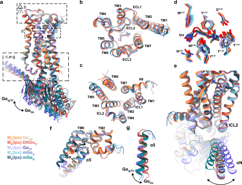
The M5 muscarinic acetylcholine receptor (M5 mAChR) represents a promising therapeutic target for neurological disorders. However, the high conservation of its orthosteric binding site poses significant challenges for drug development. While selective positive allosteric modulators (PAMs) offer a potential solution, a structural understanding of the M5 mAChR and its allosteric binding sites remains limited. Here, we present a 2.8 Å cryo-electron microscopy structure of the M5 mAChR complexed with heterotrimeric Gq protein and the agonist iperoxo, completing the active-state structural characterization of the mAChR family. To identify the binding site of M5-selective PAMs, we implement an integrated approach combining mutagenesis, pharmacological assays, structural biology, and molecular dynamics simulations. Our mutagenesis studies reveal that selective M5 PAMs bind outside previously characterized M5 mAChR allosteric sites. Subsequently, we obtain a 2.1 Å structure of M5 mAChR co-bound with acetylcholine and the selective PAM VU6007678, revealing an allosteric pocket at the extrahelical interface between transmembrane domains 3 and 4 that is confirmed through mutagenesis and simulations. These findings demonstrate the diverse mechanisms of allosteric regulation in mAChRs and highlight the value of integrating pharmacological and structural approaches to identify allosteric binding sites.


Conflict of interest statement

Competing interests: AC is a co-founder and holds equity in Septerna Inc. The remaining authors declare no competing interests.

Figures

Fig. 1
Fig. 1. Structural and functional analysis of ligand-receptor interactions at the M5 mAChR.
a ML380-ACh interaction in IP1 accumulation assay at WT M5 mAChR (left) or M5 EH4 pocket mutant (right) in CHO cells. Data points are mean ± SEM values from 7 (WT M5 mAChR) and 3 (M5 EH4 mutant) experiments performed in duplicate. An operational model of allosterism was fit to the grouped data. b Effects of the M5 mAChR mutations on the pKB of ML380. Data shown are the mean ± SEM of the affinity estimates derived from a global least-squares fit of an allosteric model to three independent experiments for all conditions, except for WT M5 mAChR (n = 7). Pharmacology parameters were shared across experiments to yield a single best estimate of each parameter and its associated standard error, as derived from the nonlinear regression algorithm. This global pooled analysis approach ensured model convergence in all instances. *, significantly different from WT, p < 0.05, one-way ANOVA, Dunnett’s post hoc test. Parameters obtained are listed in Supplementary Table 1. c M5 mAChR mutated residues shown on receptor structure. d EH4 pocket residues are shown as purple sticks. e ECV residues are shown as red sticks. f Consensus cryo-EM map of M5 mAChR-mGαq/Gβ1γ2 complex with iperoxo at 2.8 Å (FSC 0.143). Model colouring: dark blue (receptor), red (mGαq), green (Gβ1), yellow (Gγ1). g Cryo-EM density for iperoxo in orthosteric site (local refined map, contour level 0.36). h Iperoxo-orthosteric site interactions. Interaction colouring: pink (charge-charge); black (hydrogen bonds); purple (cation-π). Source data are provided as a Source Data file.
Fig. 2
Fig. 2. Structural comparison of iperoxo-bound, active state M1-M5 mAChR structures determined by cryo-EM.
a Overall view of the M1 to M5 mAChRs complexed to Gα and bound to iperoxo. Model colouring: purple (M1; PDB: 6OIJ), orange (M2; PDB: 6OIK), cyan (M3; PDB: 8E9Z), red (M4; PDB: 7TRK), dark blue (M5; PDB: 9EK0). b Extracellular view comparing ECLs and TM regions. c Intracellular view (G protein removed) comparing ICLs and TM regions. d Overlay of iperoxo and orthosteric binding site residues. e Side view comparing the αN movement of Gα. f Intracellular view comparing the α5 insertion of Gα. g Intracellular view comparing the α5 rotation of Gα. Changes are indicated by arrows.
Fig. 3
Fig. 3. Potential cryo-EM density for ML380.
a–c Local refined M5 mAChR cryo-EM map (contour level 0.3). No ML380 density was observed in a ECV or b EH4 pocket. c Potential ML380 density observed parallel to TM1/TM7 (green) and the bottom of the TM2,3,4 interface (orange). d [3H]-NMS equilibrium binding studies between [3H]-NMS, ACh and ML380 at WT M5 mAChR and M5-M2 TM chimeras. Insets show receptor cartoons (blue: M5 domains; red: M2 domains). Data points are mean ± SEM values of three duplicate experiments. An allosteric ternary complex model was fit to the data. Parameters obtained are listed in Supplementary Table 3. RMSDs (Å) of e iperoxo in orthosteric pocket and f ML380 at the potential allosteric site at the bottom of the TM2,3,4 interface from GaMD simulations of the cryo-EM structure. Three simulation replicates are shown in different colours. Source data are provided as a Source Data file.
Fig. 4
Fig. 4. High resolution structure of ACh and VU6007678 bound M5 mAChR in complex with heterotrimeric G protein.
a Consensus cryo-EM map of M5 mAChR-mGαq/Gβ1γ2 complex with ACh and VU6007678 at 2.1 Å resolution (FSC 0.143). Model colouring: light blue (receptor); red (mGαq), green (Gβ1), yellow (Gγ1), light grey (scFv16), dark grey (Nb35). b M5 mAChR model showing ACh (green) at the orthosteric site and VU6007678 (orange) at the allosteric site between TM3/TM4 and above ICL2. Cryo-EM density (local refined receptor map, contour 0.43) for c iperoxo at the orthosteric site and d VU6007678 at the allosteric site. e Comparison of orthosteric site residues. Stick colouring: light blue (ACh-bound residues), dark blue (Ipx-bound residues), light green (ACh), dark green (Ipx). f Interactions of ACh with the orthosteric binding site. Interaction colouring: pink dotted lines (charge-charge), black (hydrogen bonds), purple (cation-π); red spheres (water molecules). g 2D interaction plot of ACh with the orthosteric binding site; grey lines (hydrophobic interactions). h RMSDs (Å) of ACh relative to the starting conformation during GaMD simulations (three replicates, different colours). i VU6007678 allosteric site interactions: black (hydrogen bonds), purple (cation-π), green (π-π). j VU6007678 site comparison between the active complex (light blue) and inactive tiotropium-bound M5 (salmon, PDB:6OL9); VU6007678 (orange). k 2D VU6007678 interaction plot. Same colouring as g with green dashed lines representing π-π interactions. l RMSDs (Å) of VU6007678 relative to the starting conformation during GaMD simulations (three replicates, different colours). Source data are provided as a Source Data file.
Fig. 5
Fig. 5. Structural and functional analysis of the VU6007678 allosteric binding site.
a Comparison of VU6007678 allosteric binding site residues (stick representation) across the M1-M5 mAChRs. Colouring: purple (M1; PDB: 6OIJ), orange (M2; PDB: 6OIK), light blue (M3; PDB: 8E9Z), red (M4; PDB: 7TRK), and dark blue (M5; PDB: 9EJZ). VU6007678-ACh interaction in Trupath Gq activation assay at b WT M5 mAChR or c M5/M2 swap in CHO cells. Data points are mean ± SEM of three to eight individual experiments performed in duplicate. WT M5 mAChR n = 8, M5/M2 swap mutant n = 3. An operational model of allosterism was fitted to the grouped data to derive the key pharmacological parameters (df). Pharmacology parameter values were shared across experiments to yield a single best estimate of the mean of each parameter and its associated standard error, as derived from the nonlinear regression algorithm. This global pooled analysis approach ensured model convergence in all instances. *, significantly different from WT, p < 0.05, one-way ANOVA, Dunnett’s post hoc test. WT M5 mAChR n = 8; M5/M2 swap, F1303.52M, T1333.55A, R1343.56A, R1343.56K mutants n = 3; Y682.42F, V1233.45I, K141ICL2A, R1464.41M mutants n = 4. g VU6007678 affinity (pKB) and h log affinity cooperativity (logαβ) between ACh and VU6007876 at WT M5 mAChR and mutants from [3H]-NMS equilibrium binding studies. Data points represent mean ± SEM values of pharmacological parameters determined by fitting an allosteric ternary complex model to the grouped data, as described in (df). All pharmacology parameters (dh) are listed in Supplementary Table 4. WT M5 mAChR n = 3, F1303.52M, R1464.41M n = 4, M5/M2 swap n = 5. Time courses of distances (Å) from VU6007678 to i F1303.52, j R1464.41, k M1504.45 calculated from GaMD simulations performed with three separate replicates as indicated through different coloured traces. Source data are provided as a Source Data file.
Fig. 6
Fig. 6. Allosteric sites at the mAChRs.
a Model of the ACh-VU6007678 bound M5 mAChR showing the allosteric binding site discovered for VU6007678 (orange spheres) and the ECV allosteric site occupied by LY2119620 (salmon spheres). ACh is shown as green spheres in the orthosteric binding site. b Allosteric modulator structures overlaid on the VU allosteric site, with ligands shown as ball-and-stick models. D1:LY3154207:cholesterol in dark slate grey and slate grey, respectively (PDB:7X2F). β2AR:Cmpd-6FA in light sea green (PDB:6N48). FFAR1:AP8 in violet (PDB:5TZY). GPBAR1:INT-177 in purple (PDB:7CFN). CASR:NDT9513727 in salmon (PDB:6C1Q). C5aR:avacopan in green (PDB:6C1R). c Cryo-EM density (contour level 0.47) for lipid molecules observed at the bottom of TM2,3,4.

Update of

References

    1. Bender, A. M., Garrison, A. T. & Lindsley, C. W. The Muscarinic Acetylcholine Receptor M5: Therapeutic implications and allosteric modulation. ACS Chem. Neurosci.10, 1025–1034 (2019). - PubMed
    1. Vilaro, T. M., Palacios, J. M. & Mengod, G. Localization of m5 muscarinic receptor mRNA in rat brain examined by in situ hybridization histochemistry. Neurosci. Lett.114, 154–159 (1990). - PubMed
    1. Weiner, D. M., Levey, A. I. & Brann, M. R. Expression of muscarinic acetylcholine and dopamine receptor mRNAs in rat basal ganglia. Proc. Natl. Acad. Sci.87, 7050–7054 (1990). - PMC - PubMed
    1. Yasuda, R. P. et al. Development of antisera selective for m4 and m5 muscarinic cholinergic receptors: Distribution of m4 and m5 receptors in rat brain. Mol. Pharmacol.43, 149–157 (1993). - PubMed
    1. Foster, D. J. et al. M5 receptor activation produces opposing physiological outcomes in dopamine neurons depending on the receptor’s location. J. Neurosci.34, 3253–3262 (2014). - PMC - PubMed

LinkOut - more resources