. 2025 Jul 31;8(1):1139.
doi: 10.1038/s42003-025-08548-0.

Stoichiometric 14-3-3ζ binding promotes phospho-Tau microtubule dissociation and reduces aggregation and condensation

Affiliations

Stoichiometric 14-3-3ζ binding promotes phospho-Tau microtubule dissociation and reduces aggregation and condensation

Janine Hochmair et al. Commun Biol. .

Abstract

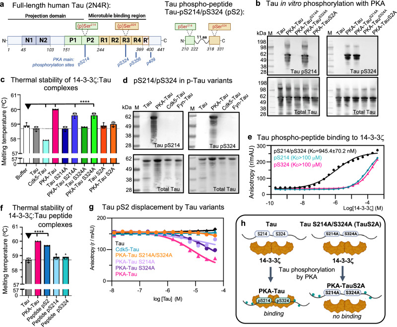
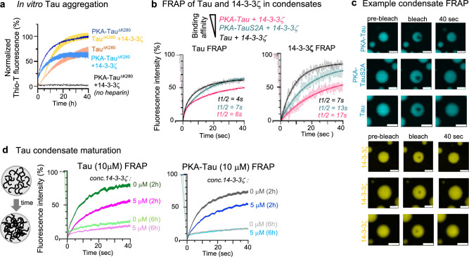
The microtubule (MT) association of protein Tau is decreased upon phosphorylation. Increased levels of phosphorylated Tau in the cytosol pose the risk of pathological aggregation, as observed in neurodegenerative diseases. We show that binding of 14-3-3ζ enhances cytosolic Tau solubility by promoting phosphorylated Tau removal from MTs, while simultaneously inhibiting Tau aggregation both directly and indirectly via suppression of condensate formation. These 14-3-3ζ activities depend on site-specific binding of 14-3-3 to Tau phosphorylated at S214 and S324. At sub-stoichiometric 14-3-3ζ concentrations, or in the presence of other 14-3-3ζ binding partners, multivalent electrostatic interactions promote Tau:14-3-3ζ co-condensation, offering a phosphorylation-independent mode of Tau-14-3-3ζ interactions. Given the high abundance of 14-3-3 proteins in the brain, 14-3-3 binding could provide efficient multi-modal chaperoning activity for Tau in the healthy brain and be important for preventing Tau aggregation in disease.


Conflict of interest statement

Competing interests: Christian Ottman and Luc Brunsveld are both co-founders of Ambagon Therapeutics. Christian Ottmann is currently employee and Luc Brunsveld is currently advisor of Ambagon Therapeutics.

Figures

Fig. 1
Fig. 1. 14-3-3ζ binding inhibits neuronal Tau aggregation.
a Representative images of primary hippocampal neurons (DIV12) transduced with AAV to express aggregating FTD-mutant eGFP-TauP301L/S320. Neurons were treated with DMSO (control), fusicoccin (50 μM), or BV02 (40 μM) for 48 h, starting 3 days after AAV transduction. Zoom-ins show examples of cell bodies with fibrillar Tau aggregates (white arrow heads) or high soluble Tau (white circles). Scale bars = 20 μm. b Quantification of Tau aggregates in neurons upon treatment with DMSO, fusicoccin (50 μM), or BV02 (10 or 40 μM). Data shown as mean ± SEM, N = 4 experimental repeats, one-way ANOVA with Tukey post-test. c Quantification of soluble eGFP-Tau (mean intensity) in the soma of neurons without aggregates. Data shown as mean ± SEM, N = 3 experimental repeats with 6–22 cells per condition, one-way ANOVA with Tukey post-test. d Representative image of fibrillar eGFP-Tau aggregate in neurons immunolabeled for MAP2 (red), 14-3-3 (white), and TaupS202/pT205 (blue; AT8 antibody). Line plot along white arrow shows no enrichment and limited colocalization of granular 14-3-3 in tangle-like Tau aggregate. Scale bar = 10 μm. e Representative image of neurons immunolabeled for 14-3-3 (cyan) and MAP2 (red). Zoom-in shows granular labeling of 14-3-3 in MAP2+ dendrites (white circles) and MAP2- axons (white arrow heads). Principle dendrites appear rich in 14-3-3. Scale bar = 10 μm. f Neurons immunolabeled for MAP2 and phospho-Tau variants indicate stronger colocalization of endogenous TaupS202/pT205 (AT8 epitope) than TaupS214/pS324 with MAP2+ microtubules (MTs). Scale bars = 10 μm. g Example fluorescence image of a neuron expressing eGFP-TauP301L/S320F, with somatodendritic MAP2-coated microtubules that are also coated with TaupS202/pT205 (AT8 antibody). Zoom-ins show that 14-3-3 granules (pink) align with TaupS202/pT205 (green) coated microtubules (compare line plot along white arrow). Scale bars = 10, 4 μm for zoom-in.
Fig. 2
Fig. 2. 14-3-3ζ binding to Tau depends on phosphorylation at S214 and S324.
a Domain structure of full-length Tau and Tau phospho-peptide, pS2. The longest human Tau isoform (2N4R, 441 aa) consists of the N-terminal projection domain with two N-terminal inserts (N1, N2), the proline-rich domain (P1 + P2), and the C-terminal microtubule binding region that includes four pseudo-repeats (R1-R4) and short sequences up- and downstream of these. Positions of phosphorylation sites on serine residues Ser214 in P2 and Ser324 in R3 are indicated. The phospho-peptide TaupS214/pS324 (pS2) has 38 aa (Tau210–222-GGGSGGGSGGG-Tau318–331). b Western blots of recombinant Tau variants in vitro phosphorylated using PKA kinase (Tau variants: wild-type Tau, TauS214A, TauS324A, TauS214A/S324A (TauS2A)) using antibodies specific for Tau phospho-residues (TaupS214 and TaupS324) and total Tau. c Thermal stability of 14-3-3ζ mixed with full-length Tau variants in their PKA-phosphorylated and non-phosphorylated forms. The higher the binding affinity, the higher the temperature needed to melt Tau:14-3-3ζ complexes. PKA-phosphorylation increases binding of Tau to 14-3-3ζ, which is reduced upon S > A mutations in TauS214A and TauS324A. Mutation of both serine residues abolishes TauS214A/S324A (=TauS2A) binding. Data shown as mean ± SD, N = 3 experimental repeats. One-way ANOVA with Tukey post-test. Significance compared to “Buffer” ( = 14-3-3ζ alone) is indicated. d Western blot of TaupS214 and TaupS324 sites in full-length Tau in vitro phosphorylated by different kinases. e Representative fluorescent anisotropy measurements for the binding of Tau phospho-peptides pS2, TaupS214, and TaupS324 to 14-3-3ζ. 14-3-3ζ was titrated into 10 nM of the respective fluorescein-labeled Tau peptide. Data shown as mean ± SD, representative experiment with N = 3 technical replicates (two more experiments shown in Supplemental Fig. S1e). f Thermal stability of 14-3-3ζ mixed with Tau phospho-peptides (pS2, TaupS214, and TaupS324) compared to full-length Tau and PKA-Tau. Data shown as mean ± SD, N = 3 independent experiments. One-way ANOVA with Tukey post-test. Significance compared to “Tau” is indicated. g Representative fluorescent anisotropy measurements for the competition between full-length phospho-Tau variants and pS2-FITC (10 nM) for the binding to 14-3-3ζ (1 μM). Data shown as mean ± SD, N = 2 technical replicates (two more experiments shown in Supplemental Fig. S1f). h Model: Full-length Tau binding to 14-3-3ζ depends on phosphorylation at S214 and S324.
Fig. 3
Fig. 3. 14-3-3ζ binding promotes Tau pS214/pS324 dissociation from microtubules.
a Western blot of the MT binding assay for Tau and PKA-Tau with and without 14-3-3ζ. b Quantification of MT binding assay. Supernatant (soluble unbound) to pellet (MT bound) S/P ratios for Tau and PKA-Tau in the presence or absence of 14-3-3. Data shown as mean ± SD, N = 4 experimental replicates, one-way ANOVA with Tukey post-test. c Model for 14-3-3ζ promoting phospho-Tau detachment from MTs. df Confocal images of in vitro MT formation with Tau variants (25 μM, 5% PEG) show Tau coating of MTs (white arrow heads) and remaining soluble Tau in solution. Non-phosphorylated Tau (d), PKA-Tau (e), and PKA-TauS2A (f). Scale bars = 20 μm, and 5 μm in zoomed insets. gi Images of MT formation in the presence of both Tau (25 μM, 5% PEG) and 14-3-3ζ (12.5 μM). When 14-3-3ζ does not bind Tau (Tau+14-3-3ζ (g) and PKA-TauS2A + 14-3-3ζR127A (i)), Tau coats MTs (white arrow heads), no free Tau is in solution, and MTs grow from condensates containing Tau, 14-3-3ζ, and tubulin (=MT asters). When 14-3-3ζ is binding Tau (PKA-Tau + 14-3-3ζ (h)), phospho-Tau is absent on MTs, and few Tau:14-3-3ζ condensates form in solution, not attached to MTs. Scale bars = 20, 5 μm in zoomed insets.
Fig. 4
Fig. 4. Stoichiometric binding of 14-3-3ζ inhibits Tau condensation.
a Representative images for the condensation of PKA-Tau, Cdk5-Tau, and Fyn-Tau (each with 2% of phosphorylation matched Tau-DyLight488) at increasing 14-3-3ζ concentrations, recorded after 2–3 h (in 25 mM HEPES, pH 7.4, 10 mM NaCl, 1 mM DTT, 5% (w/v) PEG). The graph shows quantification of condensate surface coverage (%) for PKA-Tau, Cdk5-Tau, and Fyn-Tau condensation. Data shown as mean ± SD. N = 4 experimental repeats. b, c Representative images of 10 μM PKA-Tau (a) and PKA-TauS2A (b) with increasing 14-3-3ζ concentrations (0, 1, 5, 10, 20 μM; 2% Tau-DyLight488, 2% 14-3-3ζ-DyLight647). Scale bars = 20 μm. d Quantification of condensate surface coverage (%) for 10 μM PKA-Tau and PKA-TauS214A/S324A (PKA-TauS2A) with 10 μM 14-3-3ζ or 14-3-3ζR127A. Data shown as mean ± SD. N = 3 experimental replicates. One-way ANOVA with Tukey post-test. e, f Representative images of 10 µM PKA-Tau (c) and PKA-TauS2A (d) with increasing 14-3-3ζR127A concentrations (1, 5, 10, 20 µM). Scale bars = 20 μm.
Fig. 5
Fig. 5. Tau:14-3-3ζ condensation depends on Tau availability and electrostatic interactions.
a Representative images of PKA-Tau (10 μM) with 14-3-3ζ (10 μM) and increasing concentrations of pS2 peptide (0, 5, 10, 20 μM). Scale bars = 20 μm. Quantification of condensate surface coverage (%) is shown in the bar graph. Data shown as mean ± SD. N =  two experimental replicates, three technical replicates. Data points represent individual analyzed images. One-way ANOVA with Tukey post-test. b Representative images of condensates formed by PKA-Tau (10 μM, 2% PKA-Tau-DyLight405) with 14-3-3ζ (10 μM, 2% 14-3-3ζ-DyLight650) and pS2 (20 μM, 2% FITC-pS2). Scale bars = 10 μm. c Crystal structure of 14-3-3ζ dimer (gray semi-transparent surface) in complex with pS214 and pS324 binding sites of pS2 (green rods). d Top view of pS2 binding sites around pS214 and pS324 (green rods) in complex with 14-3-3ζ (gray semi-transparent surface). The solid purple line indicates a connective unstructured 11 aa linker between the binding sites in pS2. e, f Close-up of binding grooves in 14-3-3ζ monomers 1 and 2 (gray surface) in complex with pS2 binding sites (green rods). The final 2Fo-Fc electron density map of pS2 is shown as a blue mesh (contoured at 1 s). g Surface charge distribution of 14-3-3ζ dimers mapped on crystal structure. Negatively charged areas are shown in red, positively charged ones in blue. Notably, the 14-3-3ζ binding pocket is positively charged, whereas much of the solvent-exposed surface of 14-3-3ζ is negatively charged. h Tau, PKA-Tau, and PKA-TauS2A (PKA-TaupS214A/pS324) condensation with 14-3-3ζ with increasing NaCl concentrations (1, 10, 50, 100 mM). Scale bar = 10 μm.
Fig. 6
Fig. 6. 14-3-3ζ inhibits Tau amyloid aggregation but not condensate polymerization.
a Thioflavine-T (ThioT) assay of pro-aggregant FTD-mutant TauΔK280 or PKA-TauΔK280 aggregation (triggered with heparin) in the absence and presence of 14-3-3ζ. N = 3 experimental replicates, with each 3 technical replicates. Data shown normalized to the respective conditions with maximum fluorescence (TauΔK280 + 14-3-3ζ or PKA-TauΔK280) as SD band. b FRAP of Tau variants (10 μM Tau with 2% Tau-Dylight488) and 14-3-3ζ (5 μM 14-3-3ζ with 2% 14-3-3ζ-DyLight650). N = 7-13 condensates per condition from three experiments. Data shown as mean ± SEM band. c Representative images of condensates formed from fluorescently-labeled Tau variants and 14-3-3ζ right before and after photobleaching and after 40 s of recovery. Condensates formed in 25 mM HEPES, 1 mM DTT, pH 7.4, in the presence of 5% PEG. Scale bars = 5 μm. d FRAP of “maturing” Tau and PKA-Tau condensates (10 μM; Tau with 2% Tau-Dylight488) formed in 25 mM HEPES, 1 mM DTT, pH 7.4, 5% PEG and with or without 14-3-3ζ (0 or 5 μM; with 2% 14-3-3ζ-DyLight650). Condensates were analyzed at 2 and 6 h after formation. N = 9–20 condensates per condition from three experiments. Data shown as SEM band.
Fig. 7
Fig. 7. Regulation of Tau MT binding, aggregation and condensation by 14-3-3ζ.
a 14-3-3ζ dimers form stable stoichiometric complexes with Tau pS214/pS324. This enables the efficient dissociation of Tau pS214/pS324 from MTs, which may contribute to ensure MT dynamics. In addition, the formation of 14-3-3ζ:Tau pS214/pS324 complexes prevents the aggregation of Tau in the cytosol by inhibiting phospho-Tau assembly into fibrillar aggregates and/or liquid-like condensates. In non-binding conditions, when Tau is not phosphorylated at S214 and S324, 14-3-3ζ co-condenses with Tau based on multivalent electrostatic interactions. These condensates attach to MTs, when present, and sequesters excess Tau from the solution. Whether 14-3-3ζ:Tau condensates have of cellular function remains to be clarified. b Interactions of Tau and 14-3-3 in stoichiometric binding vs. condensation conditions. In binding conditions, stoichiometric binding of Tau in the binding groove of 14-3-3ζ dimers precludes Tau condensation by depleting the pool of Tau molecules necessary to drive condensation. This is due to the occupation of TauRD and the proline-rich region in Tau—domains that drive Tau condensation—by 14-3-3ζ. In non-binding-conditions, multivalent electrostatic interactions between the 14-3-3 surface and Tau drive co-condensation. Notably, 14-3-3 dimers in complex with Tau (or other clients) can also participate in co-condensation because their surface charges are still accessible.

References

    1. Khatoon, S., Grundke-Iqbal, I. & Iqbal, K. Brain levels of microtubule-associated protein τ are elevated in Alzheimer’s disease: a radioimmuno-slot-blot assay for nanograms of the protein. J. Neurochem.59, 750–753 (1992). - PubMed
    1. Butner, K. A. & Kirschner, M. W. Tau protein binds to microtubules through a flexible array of distributed weak sites. J. Cell Biol.115, 717–730 (1991). - PMC - PubMed
    1. Wegmann, S. et al. Tau protein liquid–liquid phase separation can initiate tau aggregation. EMBO J.37, e98049 (2018). - PMC - PubMed
    1. Goedert, M. Tau filaments in neurodegenerative diseases. FEBS Lett.592, 2383–2391 (2018). - PubMed
    1. Spillantini, M. G. & Goedert, M. Tau pathology and neurodegeneration. Lancet Neurol.12, 609–622 (2013). - PubMed

LinkOut - more resources