

The efficacy of intraoperative OCT combined with foldable artificial vitreous balloon (FCVB) implantation in eyes with complex retinal detachment and silicone oil dependence
- PMID: 40745282
- PMCID: PMC12315390
- DOI: 10.1186/s12886-025-04197-3
The efficacy of intraoperative OCT combined with foldable artificial vitreous balloon (FCVB) implantation in eyes with complex retinal detachment and silicone oil dependence
Abstract
Objective: To evaluate implantation of a foldable capsular vitreous body (FCVB) in combination with intraoperative optical coherence tomography (I-OCT) for the treatment of complex retinal detachment and silicone oil-dependent eyes.
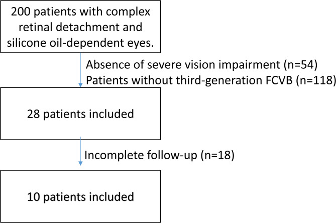
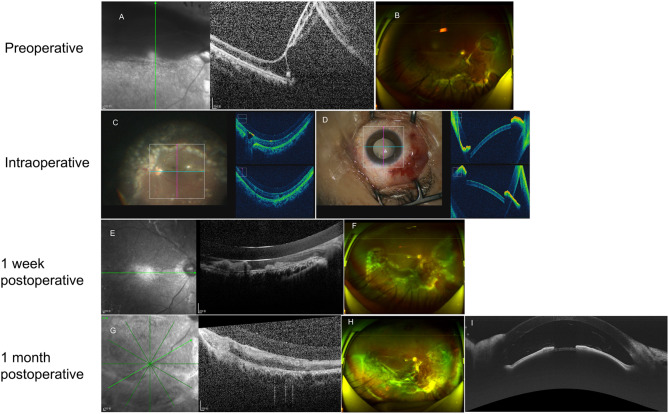
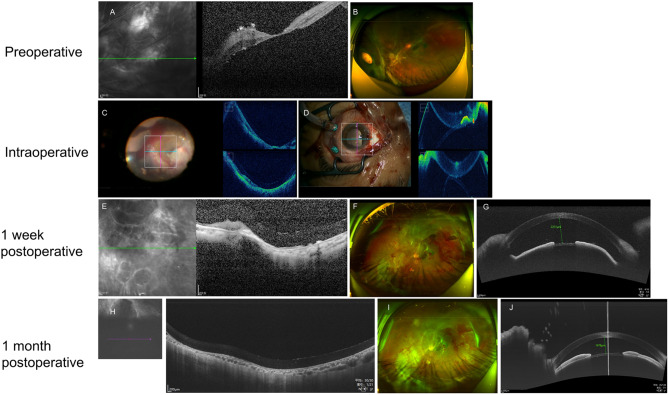
Methods: This retrospective study included 10 patients (10 eyes) who underwent third-generation FCVB implantation at the Second Affiliated Hospital of Harbin Medical University. Clinical data, including best-corrected visual acuity (BCVA) and intraocular pressure (IOP), were collected preoperatively and at 1 week, 1 month, 3 months, and 6 months postoperatively. During surgery, I-OCT was employed to dynamically monitor the silicone oil injection volume (ranging from 2.1 to 4.5 mL, based on preoperative axial length and 3D ocular reconstructions), ensuring the posterior FCVB wall adhered to the macular retina with a gap of < 100 μm, and maintaining > 500 μm clearance from the anterior capsule to the iris to preserve the posterior chamber space. Pre-, intra-, and postoperative OCT images of the anterior and posterior segments were compared.
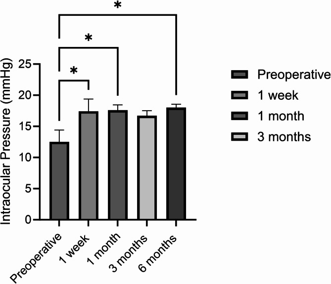
Results: All surgeries were completed successfully without intraoperative complications. I-OCT enabled real-time optimization of silicone oil volume, resulting in tight FCVB-retina adherence (< 50 μm) in 8 of 10 eyes. In 2 trauma-related cases with retinal defects, FCVB adhered directly to the sclera. BCVA showed no significant improvement at 6 months. Two cases experienced elevated IOP at 1 week post-op (34 mmHg and 22 mmHg), both of which normalized after treatment. IOP remained stable (15-22 mmHg) in all eyes at final follow-up.
Conclusion: I-OCT provided real-time quantitative feedback that allowed precise control of silicone oil injection, ensured optimal positioning of the FCVB, and reduced postoperative complications. Key intraoperative parameters included (1) real-time silicone oil volume titration, (2) gap measurement between FCVB and retina (< 100 μm), and (3) anterior chamber space evaluation (> 500 μm). The use of I-OCT significantly enhanced surgical precision and enabled individualized treatment planning.
Keywords: Complex retinal detachment; Foldable capsular vitreous body; Silicone oil-dependent eyes.
© 2025. The Author(s).
Conflict of interest statement
Declarations. Ethics approval and consent to participate: The study was approved by the Ethics Committee of The Second Affiliated Hospital of Harbin Medical University. All methods were performed in accordance with the Declarations of Helsinki, and written informed consents were signed by all participant before the study. Consent for publication: Written informed consent for publication of their clinical details and/or clinical images was obtained from the patient/parent/guardian/ relative of the patient. Conflict of interest: The authors declared that there was no conflict of interest associated with the manuscript.
Figures
Similar articles
-
Foldable capsular vitreous body indications, complications, and outcomes: a systematic review.Graefes Arch Clin Exp Ophthalmol. 2023 Aug;261(8):2103-2116. doi: 10.1007/s00417-023-05995-5. Epub 2023 Feb 16. Graefes Arch Clin Exp Ophthalmol. 2023. PMID: 36795160
-
Postoperative Outcomes of 1-Month Silicone Oil Tamponade in Rhegmatogenous Retinal Detachment: A Multicenter Study.Ophthalmic Res. 2025;68(1):333-341. doi: 10.1159/000546255. Epub 2025 May 19. Ophthalmic Res. 2025. PMID: 40388895
-
Study on the effectiveness and safety of Foldable Capsular Vitreous Body implantation.BMC Ophthalmol. 2019 Dec 18;19(1):260. doi: 10.1186/s12886-019-1268-x. BMC Ophthalmol. 2019. PMID: 31852464 Free PMC article.
-
Preliminary efficacy and safety of a silicone oil-filled foldable capsular vitreous body in the treatment of severe retinal detachment.Retina. 2012 Apr;32(4):729-41. doi: 10.1097/IAE.0b013e31822b1f80. Retina. 2012. PMID: 22105508
-
Tamponade in surgery for retinal detachment associated with proliferative vitreoretinopathy.Cochrane Database Syst Rev. 2014 Feb 14;2(2):CD006126. doi: 10.1002/14651858.CD006126.pub3. Cochrane Database Syst Rev. 2014. Update in: Cochrane Database Syst Rev. 2020 May 13;5:CD006126. doi: 10.1002/14651858.CD006126.pub4. PMID: 24532038 Free PMC article. Updated.
References
-
- Liu Y, Jiang Z, Gao Q, et al. Technical standards of a foldable capsular vitreous body in terms of mechanical, optical, and biocompatible properties. Artif Organs. 2010;34(10):836–45. 10.1111/j.1525-1594.2010.01006.x. - PubMed
-
- Lin X, Ge J, Gao Q, et al. Evaluation of the flexibility, efficacy, and safety of a foldable capsular vitreous body in the treatment of severe retinal detachment. Invest Ophthalmol Vis Sci. 2011;52(1):374–81. 10.1167/iovs.10-5869. Published 2011 Jan 21. - PubMed
-
- Zicarelli F, Staurenghi G, Invernizzi A. Anterior segment optical coherence tomography (AS-OCT) visualization of anterior vitritis. Ocul Immunol Inflamm. 2023;31(5):1101–2. 10.1080/09273948.2022.2079535. - PubMed
MeSH terms
Substances
LinkOut - more resources
Full Text Sources
Medical
Miscellaneous
