Review
. 2025 Jul 31;29(1):339.
doi: 10.1186/s13054-025-05526-0.

Assessing inspiratory drive and effort in critically ill patients at the bedside

Affiliations
Review

Assessing inspiratory drive and effort in critically ill patients at the bedside

Roberto Tonelli et al. Crit Care. .

Abstract

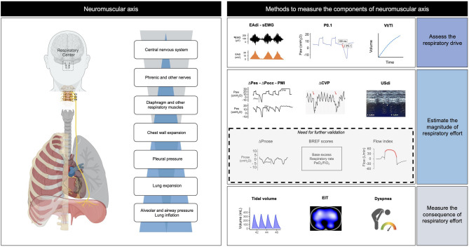
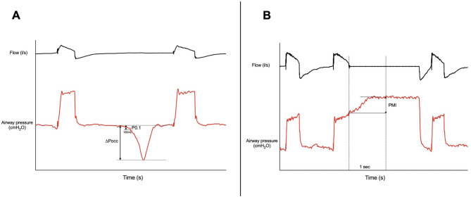
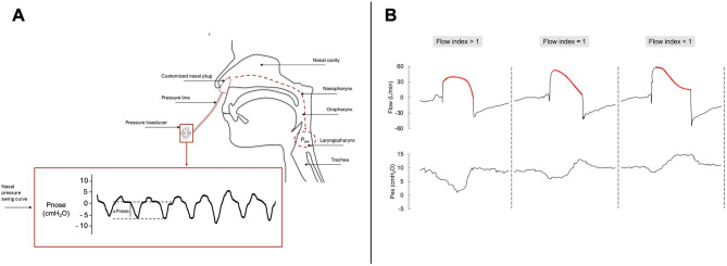
Monitoring inspiratory drive and effort may aid proper selection and setting of respiratory support in patients with acute respiratory failure (ARF), whether they are intubated or not. Although diaphragmatic electrical activity (EAdi) and esophageal manometry can be considered the reference methods for assessing respiratory drive and inspiratory effort, respectively, various alternative techniques exist, each with distinct advantages and limitations. This narrative review provides a comprehensive overview of bedside methods to assess respiratory drive and effort, with a primary focus on patients with ARF. First, EAdi and esophageal manometry are described and discussed as reference techniques. Then, alternative methods are categorized along the neuromechanical pathway from inspiratory drive to muscular effort into three groups: (1) techniques assessing the respiratory drive: airway occlusion pressure (P0.1), mean inspiratory flow (Vt/Ti) and respiratory muscle surface electromyography (sEMG); (2) techniques assessing the respiratory muscle effort: whole-breath occlusion pressure (ΔPocc), pressure-muscle index (PMI), nasal pressure swing (ΔPnose), diaphragm ultrasonography (USdi), central venous pressure swing (ΔCVP), breathing effort (BREF) models, and flow index; (3) techniques and clinical parameters assessing the consequences of effort: tidal volume (Vt), electrical impedance tomography (EIT), dyspnea. For each, we summarize the physiological rationale, measurement methodology, interpretation of results, and key limitations.

Supplementary Information: The online version contains supplementary material available at 10.1186/s13054-025-05526-0.

Keywords: Acute respiratory failure; Esophageal pressure; Inspiratory effort; Patient self-inflicted lung injury; Respiratory monitoring; Ventilator-induced diaphragm dysfunction..


Conflict of interest statement

Declarations. Ethics approval and consent to participate: Not applicable. Consent for publication: Not applicable. Competing interests: RT and EC declares patent N. 102021000007478 “APPARATO PER IL RILEVAMENTO ED IL MONITORAGGIO DELLA PRESSIONE NASALE” released on March 28th, 2023 by the Italian Ministry of Enterprises and Made in Italy. RT and EC are co-founders of IREC ltd (VAT 02959080355), (Reggio Emilia, Italy). Outside of this work, AJ has received research funding (paid to the institution) from ZonMw, Pulmotech B.V., Health~Holland, Liberate Medical, the Netherlands eScience center.

Figures

Fig. 1
Fig. 1
Techniques for bedside assessment of inspiratory drive and effort along the respiratory drive cascade. Left panel. Integrative framework linking respiratory drive and the neuromuscular axis. Transforming the central respiratory drive into effective inspiratory effort involves multiple hierarchical steps (blue pyramid). The impulse to breathe originates in the respiratory centers of the brainstem and is transmitted via the phrenic nerves to the neuromuscular junction. The activation of the respiratory muscles expands the chest wall, progressively lowering pleural pressure and expanding the lungs. As a result, alveolar and airway pressures decrease, creating a gradient relative to atmospheric pressure that drives gas into the respiratory system. As these descending processes unfold, ascending feedback mechanisms (gray pyramid) are simultaneously activated at each level. These inputs continuously relay information to the respiratory centers, enabling dynamic control of breathing. The respiratory effort can be assessed at various points along the neuromuscular axis. Right panel. Principal techniques to estimate inspiratory drive and effort at the bedside. Techniques are organized along the respiratory drive cascade and categorized according to their physiological relationship with inspiratory effort: methods assessing the respiratory drive (e.g., EAdi, P0.1, mean inspiratory flow, respiratory muscle surface EMG); methods assessing the respiratory muscle effort (e.g., ΔPes, ΔPocc, PMI, ΔPnose, USdi, ΔCVP, BREF models, and flow index); and methods evaluating the consequences of effort (e.g., tidal volume, electrical impedance tomography, dyspnea perception). Importantly, these techniques differ substantially in terms of validation status, clinical uptake, and supporting evidence. While methods such as P0.1, ΔPocc, USdi, and ΔCVP have been extensively studied and are widely adopted in both physiological research and clinical practice, others—such as ΔPnose, the flow index, and the BREF models —are promising but remain in earlier stages of development and require further clinical validation before broader implementation.ΔPes, esophageal pressure swing; ΔPocc, whole-breath occlusion pressure; P0.1, airway occlusion pressure; PMI, pressure-muscle-index; ΔPnose, nasal pressure swing; sEMG, (respiratory muscle) surface electromyography; EAdi electrical activity of the diaphragm; USdi, diaphragm ultrasonography; ΔCVP, central venous pressure swing; Vt, tidal volume; Ti, inspiratory time; EIT, electrical impedance tomography
Fig. 2
Fig. 2
Upper airway pressure trace A. How to measure P0.1 and whole-breath occlusion pressure (ΔPocc) during an end-expiratory occlusion. B. How to measure pressure-muscle-index (PMI) during an end-inspiratory occlusion. ΔPocc, whole-breath occlusion pressure; P0.1, airway occlusion pressure; PMI, pressure-muscle-index
Fig. 3
Fig. 3
A. Pnose assessment technique. This schematic illustrates the measurement of nasal pressure swing (ΔPnose) as a surrogate for inspiratory effort. The airway pressure (Paw) at the laryngopharyngeal level is influenced by inspiratory muscle activity and transmitted to the nasal cavity via the upper airway structures (nasopharynx, oropharynx, and laryngopharynx). A customized nasal plug is placed in the nostril and connected to a pressure line, which transmits pressure variations to a pressure transducer. The transducer records the nasal pressure swing curve, reflecting fluctuations in inspiratory effort over time  B. Flow Index trace. Representative flow and esophageal pressure (Pes) tracings illustrating distinct inspiratory flow decay patterns, categorized by the Flow Index. The inpiratory flow decay phase is highlighted in red. The Flow Index is a unitless parameter that describes the shape of the inspiratory flow-time curve: a value > 1 (left panel) denotes a downward-facing concavity, indicative of sustained inspiratory effort delaying flow decay; a value ≈ 1 (middle panel) reflects an approximately linear decay, consistent with a minimally active patient; a value < 1 (right panel) corresponds to an upward-facing concavity, suggesting a passive patient. A higher Flow Index has been associated with higher inspiratory effort. ΔPnose, nasal pressure swing; Paw, airway pressure; Pes, esophageal pressure
Fig. 4
Fig. 4
Schematic representation of patient-ventilator interactions during pressure support ventilation. Within a certain range of assistance, Vt matches the level set by the respiratory drive. Adjusting pressure support results in an opposite change in the inspiratory effort, with minimal changes in Vt. This specific range includes “adequate assistance”, where the effort to reach the target Vt is tolerable. This condition varies among individuals and is influenced by factors such as respiratory drive, the impedance of the respiratory system, and the muscle capacity of the patient. When pressure support exceeds this range, the inspiratory effort decreases significantly, resulting in “over-assistance”. If pressure support continues to increase, Vt will passively rise. Conversely, with “under-assistance”, the inspiratory effort to achieve the target Vt significantly increases. In extreme cases, the respiratory muscles may become unable to sustain the excessive load, causing Vt to ultimately fall short of the target. Please note that some patients may have a respiratory drive and target Vt that are too high to be achieved with a tolerable effort; even with high pressure support, under-assistance may occur. Vt, tidal volume. Pmus, respiratory muscle pressure (a measure of respiratory effort). Adapted from Docci et al. [122]
Fig. 5
Fig. 5
Key points for bedside assessment of inspiratory drive and effort. This schematic highlights essential principles to guide clinical evaluation of inspiratory drive and effort at the bedside. These include: (1) defining the clinical question and the level of precision required; (2) assessing the patient for clinical signs of low or strong effort; (3) analyzing ventilator waveforms for clues of under- or over-assistance; (4) integrating multiple monitoring techniques, as each has limitations; (5) considering trends over time, which are often more informative than isolated measurements; and (6) recognizing the gap between research data and real-life patients, particularly in underrepresented populations such as those with COPD or fibrotic lung disease. COPD, chronic obstructive pulmonary disease

Similar articles

References

    1. Battaglini D, Robba C, Ball L, et al. Noninvasive respiratory support and patient self-inflicted lung injury in COVID-19: a narrative review. Br J Anaesth. 2021;127(3):353–64. 10.1016/j.bja.2021.05.024. - PMC - PubMed
    1. Grieco DL, Maggiore SM, Roca O, et al. Non-invasive ventilatory support and high-flow nasal oxygen as first-line treatment of acute hypoxemic respiratory failure and ARDS. Intensive Care Med. 2021;47(8):851–66. 10.1007/s00134-021-06459-2. - PMC - PubMed
    1. Bellani G, Laffey JG, Pham T, et al. Noninvasive ventilation of patients with acute respiratory distress syndrome. Insights from the LUNG SAFE study. Am J Respir Crit Care Med. 2017;195(1):67–77. 10.1164/rccm.201606-1306OC. - PubMed
    1. Demoule A, Girou E, Richard JC, Taille S, Brochard L. Benefits and risks of success or failure of noninvasive ventilation. Intensive Care Med. 2006;32(11):1756–65. 10.1007/s00134-006-0324-1. - PubMed
    1. Rochwerg B, Brochard L, Elliott MW, et al. Official ERS/ATS clinical practice guidelines: noninvasive ventilation for acute respiratory failure. Eur Respir J. 2017;50(2):1602426. 10.1183/13993003.02426-2016. - PubMed

LinkOut - more resources