. 2025 Jul 31;5(1):323.
doi: 10.1038/s43856-025-01017-z.

Pantethine ameliorates dilated cardiomyopathy features in PPCS deficiency disorder in patients and cell line models

Affiliations

Pantethine ameliorates dilated cardiomyopathy features in PPCS deficiency disorder in patients and cell line models

Fangfang Zhang et al. Commun Med (Lond). .

Abstract

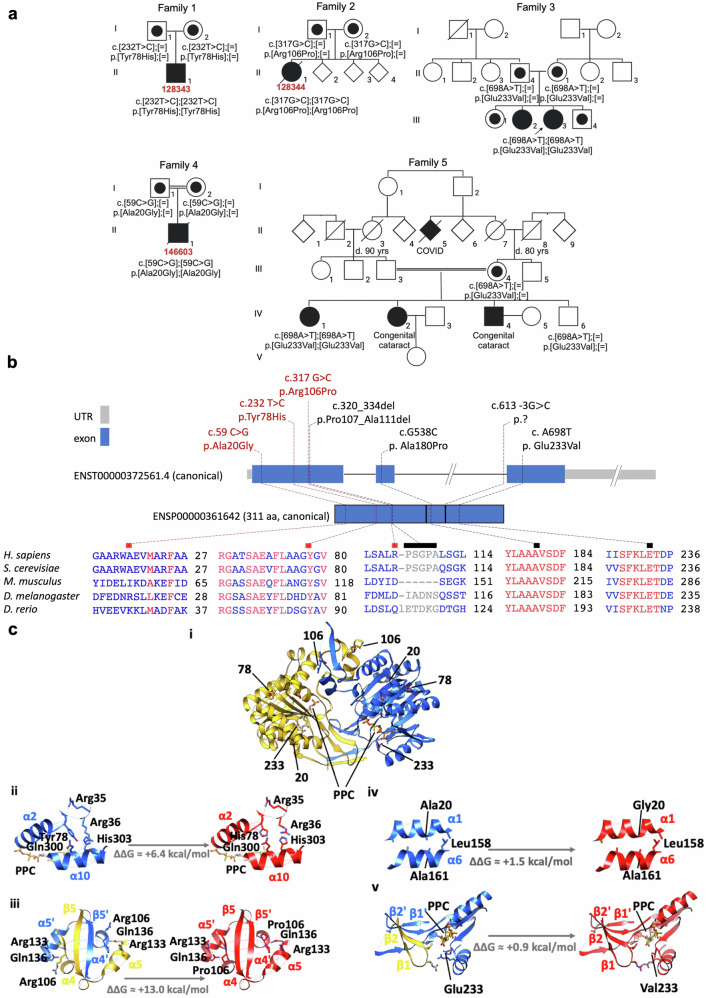
Background: PPCS deficiency disorder (PPCS DD) is an ultra-rare, autosomal recessive form of dilated cardiomyopathy (DCM) caused by pathogenic variants in PPCS, which encodes the enzyme catalyzing the second step in the coenzyme A (CoA) biosynthesis pathway. To date, only six patients worldwide have been identified.

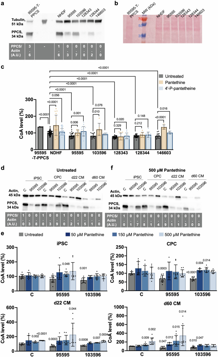
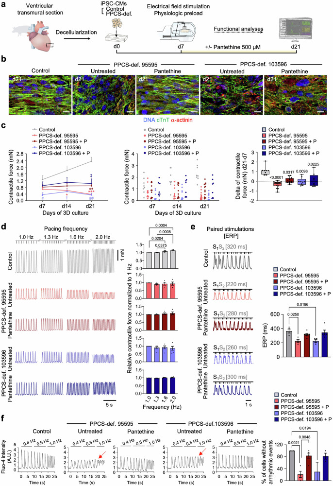
Methods: Whole-exome sequencing was performed to identify pathogenic PPCS variants in affected individuals. Protein stability was assessed by Western blotting. CoA levels were quantified using a microplate-based assay in patient-derived fibroblasts, cardiac progenitor cells, and cardiomyocytes. Functional evaluation of cardiac cells and engineered heart patches was conducted to investigate contractile performance and arrhythmogenicity. Pantethine was tested as a potential therapeutic agent both in vitro and through long-term clinical follow-up in patients.

Results: Causative PPCS variants are identified in six individuals with DCM and variable associated features, including neuromuscular and neurological symptoms. Identified variants lead to reduced PPCS protein stability and decreased cellular CoA levels. Cardiac cells exhibit impaired contractility and arrhythmias, which are partially rescued by pantethine treatment. Clinically, patients receiving pantethine show sustained improvement over time.

Conclusions: Our study expands the genetic and clinical spectrum of PPCS deficiency disorder, identifying six new cases with diverse phenotypes. Functional investigations reveal reduced CoA levels and dysfunction in patient-derived cardiac cells. Pantethine treatment shows promise in partially rescuing DCM phenotypes, both in vitro and in patients. However, complete reversal may require early intervention. These findings underscore the importance of timely diagnosis and treatment in PPCS DD. Future research should focus on optimizing pantethine supplementation and exploring additional therapies to enhance CoA levels and cardiac function in affected individuals.

Plain language summary

PPCS deficiency disorder is an extremely rare inherited disease that causes heart muscle weakness (dilated cardiomyopathy) and other symptoms. It results from changes in a gene involved in making coenzyme A (CoA), a vital molecule for cell energy. This study identified six new patients with the condition and investigated how these gene changes affect heart function. Researchers used patient cells and lab-grown heart tissues to study the disease and tested pantethine, a compound that helps increase CoA levels. They found that pantethine improved heart cell function and showed positive effects in treated patients. These results highlight the importance of early diagnosis and treatment. In the future, therapies such as pantethine could offer hope for improving heart health in affected individuals.


Conflict of interest statement

Competing interests: Sibon serves as non-compensated executive for the Spoonbill Foundation and the Stichting Lepelaar, not-for-profit organizations that may benefit from the results of this research. All other authors declare that they have no competing interests.

Figures

Fig. 1
Fig. 1. Newly identified variants occur in exon 1 of PPCS and are predicted pathogenic.
a Pedigrees of the five families with mutations in PPCS. Affected individuals are indicated with closed symbols, healthy family members with open symbols. Carriers are denoted by a dot in the center of the circle or square symbol. Fibroblast identification numbers are in red. b Localization of the newly identified (red) and already reported (black) pathogenic variants in PPCS at the gene and canonical protein level, with a zoom in the conservation of amino acid residues affected by mutations. Coloring in the sequence alignment represents the identity of amino acid residues (COBALT alignment tool). Scale gene: 100 bp = 1 cm; scale proteins: 1 cm = 33 amino acids. c Effect of Tyr78His, Arg106Pro, Ala20Gly and Glu233Val on the 3D structure of human PPCS. (i) Ribbon representation of the human PPCS dimer 3D structure (PDB code 1P9O) and side chain stick representation of Tyr78, Arg106, Ala20 and Glu233 in WT, and of His78, Pro106, Gly20 and Val233 in the thereof homology models produced with FoldX. Chain A and B of the WT protein are colored in yellow and blue, respectively, whilst residues from the mutants are colored in red. The enzymatic product analog phosphopantothenoylcystine (PPC) from the crystal structure of S. cerevisiae PPCS is shown in orange. Zoom in of the WT (left) and FoldX homology models (right) in the mutation region of (ii) Tyr78His, (iii) Arg106Pro, (iv) Ala20Gly and (v) Glu233Val highlighting residues and hydrogen bonds (dashed lines) relevant for stabilization. The ΔΔG values in kcal/mol between the WT and mutant proteins were calculated using FoldX, and account for the change in free energy for the apoprotein upon mutation, with positive values suggesting destabilization of the 3D structure and pathogenicity. The structure models in the figure were produced using UCSF Chimera.
Fig. 2
Fig. 2. Pathogenic variants in PPCS lead to reduced intracellular CoA restored through the supplementation of pantethine and 4’-P-pantetheine.
a Representative western blot analysis of fibroblasts 128343, 128344 and 146603 from newly identified patients. Fibroblasts 95595 and 103596 from previously reported patients were included as positive controls, while fibroblasts 95595 overexpressing WT PPCS (95595-T-PPCS) and NHDF from a healthy subject, were used as healthy control samples. 30 µg of total protein lysates were loaded on a 16% Tris-Glycine gel. Tubulin was used as a loading control. At the bottom of the western blot is reported the densitometric analysis of PPCS normalized to the housekeeping protein tubulin in arbitrary units (A.U.). The densitometric analysis is relative to the shown experiment. Uncrop western blot in Fig.S10. b Ponceau staining of the membrane shown in panel (a). Uncrop Ponceau staining in Fig. S11. c Intracellular levels of CoA in fibroblasts from PPCS DD patients 95595, 103596, 128343, 128344 and 146603. Fibroblasts from patient 95595 overexpressing WT PPCS (95595-T-PPCS) and NHDF were used as healthy control subjects. CoA levels in untreated NHDF were set to 100 and levels in the other fibroblast lines expressed as percent (%) of NHDF values. Measurements were performed in standard growth conditions (untreated) and in presence of 500 µM pantethine and 500 µM 4’-P-pantetheine. Results are presented as mean ± SD. Individual sample sizes are indicated for each condition (untreated: 95595-T-PPCS n = 4, NDHF n = 79, 103596 n = 13, 128343 n = 34, 128344 n = 26, 146603 n = 16; pantethine: NDHF n = 17, 95595 n = 7, 103596 n = 6, 128343 n = 9, 128344 n = 9, 146603 n = 4; 4’-P-pantetheine: NDHF n = 34, 95595 n = 17, 103596 n = 7, 128343 n = 14, 128344 n = 14, 146603 n = 4). Statistical significance relative to the untreated control was determined using an independent samples t-test, with exact p-values reported. d, e Western blot analysis of iPSCs, CPCs, d22 CMs and d60 CMs in standard growth condition (d, untreated) and after treatment with pantethine (e, 500 µM pantethine) from a healthy control subject (C) and PPCS DD patients (95595,103596). 5 µg of proteins were loaded on an 8–16% Tris-Glycine gels. Actin was used as a loading control. At the bottom of western blot is reported the densitometric analysis relative to the shown western blots. Uncrop western blot in Fig. S12. f Intracellular levels of CoA in iPSC, CPC, d22 CM and d60 CM from a healthy control subject (C) and PPCS DD patients (95595,103596). For each cell type, the levels of CoA levels in the untreated control were set to 100 and levels in the other cell lines express as percent (%) of control value. Measurements were performed in standard growth conditions (untreated) and in presence of 50, 150 and 500 µM pantethine. Results are presented as mean ± SD. Individual sample sizes are indicated for each condition (iPSC untreated: C n = 10, 95595 n = 11, 103596 n = 10; iPSC 50 µM pantethine: C n = 7, 95595 n = 7, 103596 n = 7; iPSC 150 µM pantethine: C n = 9, 95595 n = 8, 103596 n = 9; iPSC 500 µM pantethine C n = 9, 95595 n = 9, 103596 n = 8; CPC untreated: C n = 8, 95595 n = 11, 103596 n = 9; CPC 50 µM pantethine: C n = 7, 95595 n = 6, 103596 n = 6; CPC 150 µM pantethine: C n = 9, 95595 n = 9, 103596 n = 7; CPC 500 µM pantethine C n = 9, 95595 n = 8, 103596 n = 7; d22 CM untreated: C n = 9, 95595 n = 10, 103596 n = 7; d22 CM 50 µM pantethine: C n = 6, 95595 n = 7, 103596 n = 4; d22 CM 150 µM pantethine: C n = 7, 95595 n = 8, 103596 n = 6; d22 CM 500 µM pantethine C n = 7, 95595 n = 9, 103596 n = 6; d60 CM untreated: C n = 7, 95595 n = 6, 103596 n = 9; d60 CM 50 µM pantethine: C n = 7, 95595 n = 7, 103596 n = 4; d60 CM 150 µM pantethine: C n = 8, 95595 n = 9, 103596 n = 6; d60 CM 500 µM pantethine C n = 9, 95595 n = 9, 103596 n = 6). Statistical significance relative to the control was determined using an independent samples t-test, with exact p-values reported. Uncrop western blot in Fig. S13.
Fig. 3
Fig. 3. Pantethine ameliorates PPCS DD disease phenotypes in patient iPSC-derived 3D heart patches.
a Heart patches were generated by seeding d15 CMs from control and PPCS deficient iPSCs (patients 95595 and 103596) onto decellularized extracellular matrix from porcine ventricular myocardium. After 7 days, recellularized patches were transferred to biomimetic chambers allowing continuous electromechanical stimulation; from this day on PPCS deficient patches were either untreated or treated with 500 µM pantethine until day 21 of 3D culture. b Immunofluorescence staining of cTnT and α-actinin at day 21 in control patches and PPCS deficient patches with or without pantethine. Scale bars = 10 µm. Images are representative of n = 3 patches per group. c (Left and middle) Contraction force of control patches and PPCS deficient patches with or without pantethine (P) on days 7, 14, and 21 of 3D culture. Mean ± SEM (left) and individual data points (middle), control (two lines): n = 8 patches; PPCS deficient 95595: untreated n = 15, + P n = 13 patches; PPCS deficient 103596: untreated n = 9, + P n = 8 patches. Two-way ANOVA with Tukey’s multiple comparisons test, d7: p = 0.0420 (#) PPCS deficient 103596 untreated vs control; d14: p = 0.0179 (*) PPCS deficient 95595 untreated vs control, p = 0.0138 (†) PPCS deficient 103596 untreated vs control; d21: p = 0.0003 (***) PPCS deficient 95595 untreated vs control, p = 0.0020 (**) PPCS deficient 95595 + P vs control; p = 0.0002 (##) PPCS deficient 103596 untreated vs control, p = 0.0230 (‡) PPCS deficient 103596 + P vs control. The source data are provided in the Supplementary Data 6. (Right) Corresponding delta of contractile force d21-d7. Box plots show all data points and indicate the median and 25th and 75th percentiles, with whiskers extending to the min and max values. Kruskal-Wallis test with Dunn’s multiple comparisons test, p-values vs control. d (Left) Representative traces of the contractile force of control patches and PPCS deficient patches with or without pantethine placed under increasing pacing frequencies (1.0, 1.3, 1.6, and 2.0 Hz) to assess their force-frequency relationship (FFR) on day 21 of 3D culture. (Right) Contractile force relative to 1 Hz, indicated as mean ± SEM. Control (two lines): n = 5 patches; PPCS deficient 95595: untreated n = 4, + P n = 4 patches; PPCS deficient 103596: untreated n = 5, + P n = 4 patches. Two-way ANOVA with Tukey’s multiple comparisons test. e (Left) Representative traces of the contractile force of control patches and PPCS deficient patches with or without pantethine submitted to paired stimulations at different intervals to determine their effective refractory period (ERP) on day 21 of 3D culture. (Right) ERP indicated as mean ± SEM. Control (two lines): n = 4 patches; PPCS deficient 95595: untreated n = 4, + P n = 7 patches; PPCS deficient 103596: untreated n = 4, + P n = 5 patches. Kruskal-Wallis test with Dunn’s multiple comparisons test. f (Left) Representative single-cell Ca2+ transients in control iPSC-CMs and PPCS deficient iPSC-CMs (patients 95595 and 103596) within patches with or without treatment with 500 µM pantethine, placed under 0.4, 0.5, and 1 Hz pacing frequencies. Red arrows indicate examples of arrhythmic events. (Right) Percentage of CMs without arrhythmic events, indicated as mean ± SEM; control (two lines): n = 3 independent differentiations; PPCS deficient 95595: untreated n = 4, + P n = 5 independent differentiations; PPCS deficient 103596: untreated n = 2, + P n = 3 independent differentiations. One-way ANOVA.

Similar articles

References

    1. Daugherty, M. et al. Complete Reconstitution of the Human Coenzyme A Biosynthetic Pathway via Comparative Genomics. J. Biol. Chem.277, 21431–21439 (2002). - PubMed
    1. Iuso, A. et al. Mutations in PPCS, Encoding Phosphopantothenoylcysteine Synthetase, Cause Autosomal-Recessive Dilated Cardiomyopathy. Am. J. Hum. Genet.102, 1018–1030 (2018). - PMC - PubMed
    1. Lok, A. et al. Novel phosphopantothenoylcysteine synthetase (PPCS) mutations with prominent neuromuscular features: Expanding the phenotypical spectrum of PPCS-related disorders. Am. J. Med. Genet. A. (2022) 10.1002/ajmg.a.62848. - PubMed
    1. Bravo‐Alonso, I. et al. Pathogenic variants of the coenzyme A biosynthesis‐associated enzyme phosphopantothenoylcysteine decarboxylase cause autosomal‐recessive dilated cardiomyopathy. J. Inherit. Metab. Dis. 12584 (2023) 10.1002/jimd.12584. - PubMed
    1. Bosveld, F. et al. De novo CoA biosynthesis is required to maintain DNA integrity during development of the Drosophila nervous system. Hum. Mol. Genet.17, 2058–2069 (2008). - PubMed

LinkOut - more resources