

Advanced multi-label brain hemorrhage segmentation using an attention-based residual U-Net model
- PMID: 40745607
- PMCID: PMC12315353
- DOI: 10.1186/s12911-025-03131-3
Advanced multi-label brain hemorrhage segmentation using an attention-based residual U-Net model
Abstract
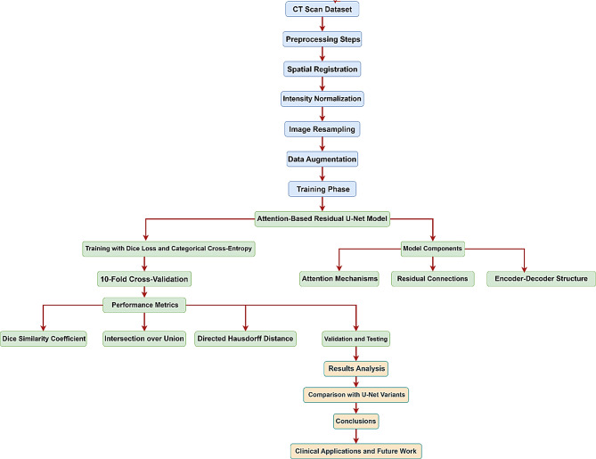
Objective: This study aimed to develop and assess an advanced Attention-Based Residual U-Net (ResUNet) model for accurately segmenting different types of brain hemorrhages from CT images. The goal was to overcome the limitations of manual segmentation and current automated methods regarding precision and generalizability.
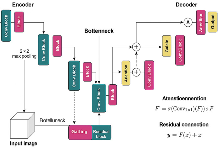
Materials and methods: A dataset of 1,347 patient CT scans was collected retrospectively, covering six types of hemorrhages: subarachnoid hemorrhage (SAH, 231 cases), subdural hematoma (SDH, 198 cases), epidural hematoma (EDH, 236 cases), cerebral contusion (CC, 230 cases), intraventricular hemorrhage (IVH, 188 cases), and intracerebral hemorrhage (ICH, 264 cases). The dataset was divided into 80% for training using a 10-fold cross-validation approach and 20% for testing. All CT scans were standardized to a common anatomical space, and intensity normalization was applied for uniformity. The ResUNet model included attention mechanisms to enhance focus on important features and residual connections to support stable learning and efficient gradient flow. Model performance was assessed using the Dice Similarity Coefficient (DSC), Intersection over Union (IoU), and directed Hausdorff distance (dHD).
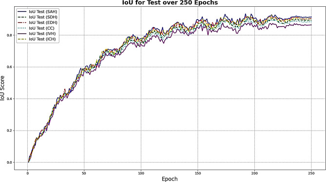
Results: The ResUNet model showed excellent performance during both training and testing. On training data, the model achieved DSC scores of 95 ± 1.2 for SAH, 94 ± 1.4 for SDH, 93 ± 1.5 for EDH, 91 ± 1.4 for CC, 89 ± 1.6 for IVH, and 93 ± 2.4 for ICH. IoU values ranged from 88 to 93, with dHD between 2.1- and 2.7-mm. Testing results confirmed strong generalization, with DSC scores of 93 for SAH, 93 for SDH, 92 for EDH, 90 for CC, 88 for IVH, and 92 for ICH. IoU values were also high, indicating precise segmentation and minimal boundary errors.
Conclusions: The ResUNet model outperformed standard U-Net variants, achieving higher multi-label segmentation accuracy. This makes it a valuable tool for clinical applications that require fast and reliable brain hemorrhage analysis. Future research could investigate semi-supervised techniques and 3D segmentation to further enhance clinical use.
Clinical trial number: Not applicable.
Keywords: Attention-based residual U-Net; Brain hemorrhage; CT; Deep learning; Medical imaging; Segmentation.
© 2025. The Author(s).
Conflict of interest statement
Declarations. Ethics approval and consent to participate: The study adhered to the Declaration of Helsinkiin in compliance with the Declaration of Helsinki. The need for ethics approval was waived by an ethics committee from the Department of General rehabilitation, The Second Affiliated Hospital and Yuying Children’s Hospital of Wenzhou Medical University, China (Reference Number: 1412-1122E). In terms of “Consent to Participate”, all participants were fully informed about the study’s purpose, procedures, and their right to withdraw at any time without penalty. Informed consent was obtained from all individual participants included in the study. For participants under 16 years of age, written informed consent was obtained from their parents or legal guardians. Consent for publication: Not applicable. Competing interests: The authors declare no competing interests.
Figures


References
-
- Hu P, Zhou H, Yan T, Miu H, Xiao F, Zhu X, et al. Deep learning-assisted identification and quantification of aneurysmal subarachnoid hemorrhage in non-contrast CT scans: development and external validation of hybrid 2D/3D UNet. NeuroImage. 2023;279:120321. - PubMed
MeSH terms
LinkOut - more resources
Full Text Sources
Medical
